

Minimally Invasive Surgical Repair of a Partial Atrioventricular Canal Defect in a 20-Year-Old Patient-A Case Report and Review of Literature
- PMID: 36286304
- PMCID: PMC9604241
- DOI: 10.3390/jcdd9100352
Minimally Invasive Surgical Repair of a Partial Atrioventricular Canal Defect in a 20-Year-Old Patient-A Case Report and Review of Literature
Abstract
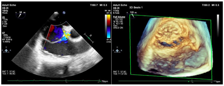
The association of an ostium primum-type defect with a cleft anterior mitral valve is known in the medical literature as the partial form of an atrioventricular canal. We present a case report about a 20-year-old woman with minimal symptomatology that discovered her pathology on routine echocardiography. Today, surgical operation remains the gold standard in such pathologies, especially mandatory when there is important valvular regurgitation and left-to-right shunt. Currently living in the era of fast and good cosmetic outcomes, minimally invasive and endovascular approaches should be developed and more often practiced. This scientific presentation is the first step in showing our department steps in performing minimally invasive surgeries as a routine.
Keywords: atrioventricular defect; minimally invasive surgery; mitral valve; valvular regurgitation.
Conflict of interest statement
The authors declare no conflict of interest.
Figures


References
-
- Carpentier A. Pediatric Cardiology. Churchill Livingstone; London, UK: 1978. Surgical anatomy and management of the mitral component of atrioventricular canal defects; pp. 477–486.
-
- Iliuta L., Rac-Albu M. Predictors and late incidence of persistent or recurrent heart failure after aortic valve replacement for aortic stenosis compared with aortic regurgitation. Eur. Heart J. 2014;35:58.
Publication types
LinkOut - more resources
Full Text Sources
