. 2022 Oct;119(4):604-615.
doi: 10.36660/abc.20210708.

Vascular Aging and Arterial Stiffness

[Article in English, Portuguese]
Affiliations

Vascular Aging and Arterial Stiffness

[Article in English, Portuguese]
Adriana Camargo Oliveira et al. Arq Bras Cardiol. 2022 Oct.

Abstract

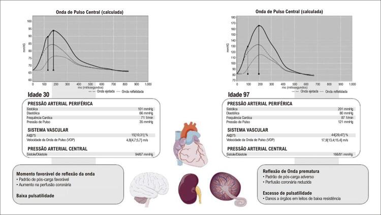
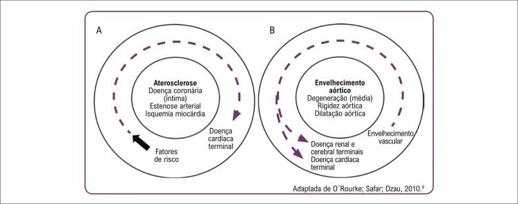
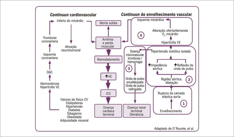
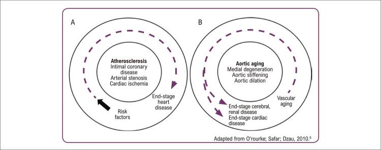
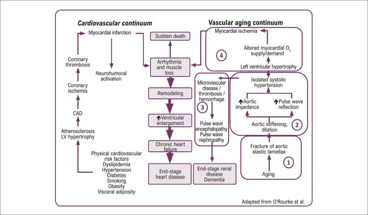
Biological aging occurs as a result of the interaction between genetics, chronological age and external factors. It is the basis for new concepts of vascular aging, whose progression is determined by the difference between biological and chronological age. From the structural point of view, the effects of vascular aging are more evident in the tunica media of large elastic arteries, marked by increased arterial stiffness, lumen dilation and wall thickness. These effects are described in the continuum of cardiovascular aging (proposed by Dzau in 2010), in which the progressive steps of microvasculature lesions of the heart, kidney and brain are initiated from the aging process. The increase of arterial stiffness can be detected by several non-invasive methods. Cardiovascular events have been traditionally described using scores that combine conventional risk factors for atherosclerosis. In the classic cardiovascular continuum (Dzau, 2006), to determine the exact contribution of each risk factor is challenging; however, since arterial stiffness reflects both early and cumulative damage of these cardiovascular risk factors, it is an indicator of the actual damage to the arterial wall. This article provides a general overview of pathophysiological mechanisms, arterial structural changes, and hemodynamic consequences of arterial stiffness; non-invasive methods for the assessment of arterial stiffness and of central blood pressure; the cardiovascular aging continuum, and the application of arterial stiffness in cardiovascular risk stratification.

O envelhecimento biológico é reflexo da interação entre genética, idade cronológica e fatores externos; é a base para novos conceitos em envelhecimento vascular, cuja progressão é determinada pela diferença entre idade biológica e cronológica. Do ponto de vista estrutural, os efeitos do envelhecimento vascular são mais evidentes na camada média das grandes artérias elásticas e resultam em aumento da rigidez arterial, da dilatação do lúmen e da espessura da parede. Esses efeitos são descritos no continuum de envelhecimento cardiovascular (proposto por Dzau em 2010) em que as etapas progressivas de lesões da microvasculatura de coração, rins e cérebro, têm início a partir do processo de envelhecimento. O aumento da rigidez arterial pode ser verificado de forma não invasiva por vários métodos. Os eventos cardiovasculares têm sido tradicionalmente previstos utilizando escores que combinam fatores de risco convencionais para aterosclerose. No continuum cardiovascular clássico (Dzau, 2006), é desafiador avaliar o peso exato da contribuição de cada fator de risco; entretanto, por refletir o dano precoce e cumulativo desses fatores de riscos cardiovascular, a rigidez arterial reflete o verdadeiro dano à parede arterial. Este artigo fornece uma visão geral dos mecanismos da fisiopatogenia, alterações estruturais das artérias e consequências hemodinâmicas do envelhecimento arterial; métodos não invasivos para a avaliação da rigidez arterial e da medida central da pressão arterial; o continuum de envelhecimento cardiovascular, e aplicação do conceito de rigidez arterial na estratificação de risco cardiovascular.


Conflict of interest statement

Potencial conflito de interesse

Não há conflito com o presente artigo

Figures

Figura 1
Figura 1. A rigidez arterial nas grandes artérias. Em adultos jovens saudáveis, uma aorta complacente (esquerda): 1) protege efetivamente o excesso de pulsatilidade causado pela ejeção ventricular esquerda intermitente; e 2) exibe uma velocidade de onda de pulso (VOP) mais lenta, permitindo que as ondas de pulso refletidas cheguem ao coração durante a diástole, aumentando a pressão de perfusão coronariana diastólica, mas não a pós-carga. Vários fatores, como envelhecimento e estilo de vida, aumentam a rigidez da parede aórtica, o que leva a várias consequências hemodinâmicas adversas. O enrijecimento aórtico leva ao aumento da impedância da raiz aórtica, com consequente aumento da amplitude da onda e chegada precoce de ondas refletidas ao coração. Essas alterações hemodinâmicas resultam em padrões adversos de carga pulsátil para o ventrículo esquerdo na sístole e redução da pressão de perfusão coronariana na diástole, em última análise, promovendo remodelamento miocárdico, disfunção e redução da reserva de perfusão (mesmo na ausência de doença coronariana epicárdica). Esse padrão hemodinâmico inverso também resulta em pulsatilidade excessiva na aorta, que é transmitida preferencialmente para leitos vasculares de baixa resistência (como rim, placenta e cérebro), pois nesses órgãos a pressão microvascular está mais diretamente associada às flutuações da pressão da arterial aórtica. Fonte: autores.
Figura 2
Figura 2. Medida da velocidade da onda de pulso carotídeo-femoral com o método “pé-a-pé”. Medida da velocidade da onda de pulso carotídeo-femoral com o método pé-pé. As formas de onda são geralmente obtidas por via transcutânea na artéria carótida comum direita e na artéria femoral direita. O atraso de tempo (Δt ou tempo de trânsito) é medido entre os pés das duas formas de onda. A distância (ΔL) percorrida pelas ondas é geralmente a distância da superfície entre os dois locais de registro, ou seja, a artéria carótida comum e a artéria femoral comum. A Velocidade de Onda de Pulso (VOP) é calculada como VOP = 0,8 × ΔL (m) / Δt (s). Fonte: autores
Figura 3
Figura 3. A forma da onda de pressão carotídea registrada por tonometria de aplanação. O fenômeno da reflexão da onda pode ser quantificado por meio do índice de aumento (AIx) - definido como a diferença entre o segundo (P2) e o primeiro (P1) picos sistólicos (P2 - P1 = PA, ou seja, pressão de aumento) expresso como uma porcentagem de PP (pressão de pulso), AIx = PA / PP. Fonte: autores.
Figura 4
Figura 4. Comparação entre o continuum cardiovascular clássico (A) e o continuum do envelhecimento cardiovascular (B). Fonte: Barroso; Barbosa; Mota-Gomes, 2020.
Figura 5
Figura 5. Associação do continuum cardiovascular clássico com o Continuum do envelhecimento cardiovascular; DAC: doença arterial coronariana; VE: ventrículo esquerdo; ICC: insuficiência cardíaca crônica. Fonte: Barroso; Barbosa; Mota-Gomes, 2020.
Figure 1
Figure 1. Arterial stiffness in large arteries. In a young health individual, a compliant aorta (left): 1) protects excess pulsatility caused by the intermittent left ventricular ejection and 2) exhibits a slow pulse wave velocity (PWV), allowing that reflected waves arrive to the heart during diastole, increasing diastolic coronary perfusion pressure but not after-load. Factors like aging and lifestyle increase aortic wall stiffness, leading to adverse hemodynamic consequences. Aortic stiffness leads to a rise in aortic root impedance, with consequent increase in forward wave amplitude and earlier arrival of the reflected waves to the heart. These hemodynamic changes result in adverse patterns of pulsatile load to the left ventricle during systole and a reduction in perfusion reserve (even in the absence of epicardial coronary disease). This inverse hemodynamic pattern also causes excess pulsatility in the aorta, which is preferably transferred to low-resistance beds, such as the kidney, placenta, and brain. In these organs, microvascular pressure is more directly associated with fluctuations in aortic pressure. AIx@75: augmentation index adjusted at heart rate. Source: the authors.
Figure 2
Figure 2. Measurement of carotid-femoral pulse wave velocity with the foot-to- foot method. Measurement of carotid-femoral pulse wave velocity with the foot-to-foot method. The waveforms are usually obtained transcutaneously, at the right common carotid artery and the right femoral artery. The time delay (Δt, or transit time) is measured between the feet of the two waveforms (Figure 1). The distance (ΔL) covered by the waves is usually the surface distance between two recording sites, i.e., the common carotid and the femoral artery. Pulse wave velocity (PWV) is calculated as PWV = 0.8 x ΔL (m)/Δt (s). Source: the authors.
Figure 3
Figure 3. Carotid pressure waveform recorded by applanation tonometry. The wave reflection phenomenon can be quantified by the augmentation index (AIx), defined as the difference between the first (P1) and the second (P2) systolic peaks (P2 - P1 = AP, i.e., augmentation pressure), expressed as a percentage of pulse pressure (PP), AIx = AP / PP. Source: the authors.
Figure 4
Figure 4. Comparison between classic cardiovascular continuum (A) and aging cardiovascular continuum (B). Source: Barroso; Barbosa; Mota-Gomes, 2020.
Figure 5
Figure 5. Association between classic cardiovascular continuum and aging cardiovascular continuum. LV: left ventricular; CAD: coronary artery disease; VE: ventricular enlargement. Source: Barroso; Barbosa; Mota-Gomes, 2020.

References

    1. Hamczyk MR, Nevado RM, Barettino A, Fuster V, Andrés V. Biological Versus Chronological Aging: JACC Focus Seminar. 10.1016/j.jacc.2019.11.062.J Am Coll Cardiol. 2020 Mar 3;75(8):919–930. - PubMed
    1. Wang M, Monticone RE, McGraw KR. Proinflammatory Arterial Stiffness Syndrome: A Signature of Large Arterial Aging. 10.1159/000490244.J Vasc Res. 2018;55(4):210–223. - PMC - PubMed
    1. Barroso W, Barbosa E, Mota-Gomes A. Rigidez Arterial e Hemodinâmica Central: Do Endotélio à Camada Média. São Paulo: Athos Mais Editora; 2020.
    1. Cunha PG, Boutouyrie P, Nilsson PM, Laurent S. Early Vascular Ageing (EVA): Definitions and Clinical Applicability. 10.2174/1573402113666170413094319.Curr Hypertens Rev. 2017;13(1):8–15. - PubMed
    1. Nilsson PM, Boutouyrie P, Cunha P, Kotsis V, Narkiewicz K, Parati G, et al. Early Vascular Ageing in Translation: From Laboratory Investigations to Clinical Applications in Cardiovascular Prevention. 10.1097/HJH.0b013e328361e4bd.J Hypertens. 2013;31(8):1517–1526. - PubMed