. 2023 Mar;68(3):267-278.
doi: 10.1165/rcmb.2021-0508OC.

Microbial-induced Redox Imbalance in the Neonatal Lung Is Ameliorated by Live Biotherapeutics

Affiliations

Microbial-induced Redox Imbalance in the Neonatal Lung Is Ameliorated by Live Biotherapeutics

Amelia E Freeman et al. Am J Respir Cell Mol Biol. 2023 Mar.

Abstract

Bronchopulmonary dysplasia (BPD) is a common lung disease of premature infants. Hyperoxia exposure and microbial dysbiosis are contributors to BPD development. However, the mechanisms linking pulmonary microbial dysbiosis to worsening lung injury are unknown. Nrf2 (nuclear factor erythroid 2-related factor 2) is a transcription factor that regulates oxidative stress responses and modulates hyperoxia-induced lung injury. We hypothesized that airway dysbiosis would attenuate Nrf2-dependent antioxidant function, resulting in a more severe phenotype of BPD. Here, we show that preterm infants with a Gammaproteobacteria-predominant dysbiosis have increased endotoxin in tracheal aspirates, and mice monocolonized with the representative Gammaproteobacteria Escherichia coli show increased tissue damage compared with germ-free (GF) control mice. Furthermore, we show Nrf2-deficient mice have worse lung structure and function after exposure to hyperoxia when the airway microbiome is augmented with E. coli. To confirm the disease-initiating potential of airway dysbiosis, we developed a novel humanized mouse model by colonizing GF mice with tracheal aspirates from human infants with or without severe BPD, producing gnotobiotic mice with BPD-associated and non-BPD-associated lung microbiomes. After hyperoxia exposure, BPD-associated mice demonstrated a more severe BPD phenotype and increased expression of Nrf2-regulated genes, compared with GF and non-BPD-associated mice. Furthermore, augmenting Nrf2-mediated antioxidant activity by supporting colonization with Lactobacillus species improved dysbiotic-augmented lung injury. Our results demonstrate that a lack of protective pulmonary microbiome signature attenuates an Nrf2-mediated antioxidant response, which is augmented by a respiratory probiotic blend. We anticipate antioxidant pathways will be major targets of future microbiome-based therapeutics for respiratory disease.

Keywords: antioxidant; humanized microbiome; microbiota; neonatal lung injury; redox.


Figures

Figure 1.
Figure 1.
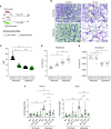
Gammaproteobacteria-predominant dysbiosis transplanted from human infants with severe bronchopulmonary dysplasia (BPD) is sufficient to augment lung injury in humanized gnotobiotic mice. (A) Tracheal aspirates from human preterm infants with or without severe BPD were analyzed for endotoxin concentration and saved for the creation of gnotobiotic mice (schematic). (B) Preterm infants with severe BPD have higher endotoxin concentrations in tracheal aspirate fluid than postmenstrual age–matched control infants without disease. (C) Humanized BPD (hBPD) and preterm-associated (hPT) mice were created by transplantation of the airway microbiome from infants with severe BPD and control infants without disease into germ-free (GF) mice. (D) Microbiome analysis demonstrating preserved differences in hBPD mice that model differences seen in human infants with severe BPD. (E) Representative photomicrographs of hematoxylin and eosin–stained sections of lungs from hBPD, hPT, and GF mice exposed to normoxia or hyperoxia, showing decreased radial alveolar counts (RACs) and increased mean linear intercept in hBPD mice compared with either control (hPT or GF) and a further augmentation of lung injury in hBPD mice exposed to hyperoxia. Scale bars, 100 μm. (F) In hyperoxia, hBPD mice exhibit more restrictive and less compliant pulmonary mechanics than hPT or GF mice. Data are presented as mean ± SEM and representative of n = 4–10 mice per experimental group. *P < 0.05, **P < 0.01, ***P < 0.001, and ****P < 0.0001 by two-way ANOVA with Tukey’s post hoc test. Schematics created using BioRender.com. P = postnatal day; PCoA = principal coordinate analysis; RDA = redundancy analysis.
Figure 1.
Figure 1.
Gammaproteobacteria-predominant dysbiosis transplanted from human infants with severe bronchopulmonary dysplasia (BPD) is sufficient to augment lung injury in humanized gnotobiotic mice. (A) Tracheal aspirates from human preterm infants with or without severe BPD were analyzed for endotoxin concentration and saved for the creation of gnotobiotic mice (schematic). (B) Preterm infants with severe BPD have higher endotoxin concentrations in tracheal aspirate fluid than postmenstrual age–matched control infants without disease. (C) Humanized BPD (hBPD) and preterm-associated (hPT) mice were created by transplantation of the airway microbiome from infants with severe BPD and control infants without disease into germ-free (GF) mice. (D) Microbiome analysis demonstrating preserved differences in hBPD mice that model differences seen in human infants with severe BPD. (E) Representative photomicrographs of hematoxylin and eosin–stained sections of lungs from hBPD, hPT, and GF mice exposed to normoxia or hyperoxia, showing decreased radial alveolar counts (RACs) and increased mean linear intercept in hBPD mice compared with either control (hPT or GF) and a further augmentation of lung injury in hBPD mice exposed to hyperoxia. Scale bars, 100 μm. (F) In hyperoxia, hBPD mice exhibit more restrictive and less compliant pulmonary mechanics than hPT or GF mice. Data are presented as mean ± SEM and representative of n = 4–10 mice per experimental group. *P < 0.05, **P < 0.01, ***P < 0.001, and ****P < 0.0001 by two-way ANOVA with Tukey’s post hoc test. Schematics created using BioRender.com. P = postnatal day; PCoA = principal coordinate analysis; RDA = redundancy analysis.
Figure 2.
Figure 2.
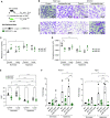
Monocolonization with Escherichia coli exacerbates lung injury. (A) Newborn GF mice were monocolonized with E. coli before randomization to normoxia or hyperoxia together with GF control mice. (B) Photomicrographs of hematoxylin and eosin–stained sections of lungs revealed alveolar hypoplasia in monocolonized mice with hyperoxia exposure. Scale bars, 100 μm. (C) RACs were significantly decreased by the combined exposure of monocolonization and hyperoxia. (D) Monocolonized mice exposed to hyperoxia had increased resistance. (E) Monocolonized mice exposed to hyperoxia had decreased compliance. (F) GF mice show significantly less Heme Oxygenase 1 (Hmox1) and NAD(P)H Quinone Dehydrogenase 1 (Nqo1) activity than wild-type (WT) control mice in hyperoxia. Fold change is in reference to GF control mice at 21% FiO2. Data are presented as mean ± SEM and representative of n = 4–8 mice per experimental group. *P < 0.05, **P < 0.01, ***P < 0.001, and ****P < 0.0001 by two-way ANOVA with Tukey’s post hoc test. Schematic created using BioRender.com.
Figure 3.
Figure 3.
Nrf2 (nuclear factor–erythroid factor 2–related factor 2) deficiency exacerbates lung injury in mice with E. coli microbiome augmentation. (A) Newborn Nrf2−/− (KO) or WT C57BL/6 control mice were intranasally instilled with E. coli before randomization to normoxia or hyperoxia. (B) Photomicrographs of hematoxylin and eosin–stained sections of lungs revealed alveolar hypoplasia in Nrf2−/− that is augmented by E. coli and hyperoxia exposure. Scale bars, 100 μm. (C) RACs were significantly decreased by the combined exposure of E. coli and hyperoxia in Nrf2−/− mice. (D) Nrf2−/− mice exposed to hyperoxia and E. coli had decreased compliance compared with WT control mice. (E) Nrf2−/− mice exposed to hyperoxia and E. coli had increased resistance compared with WT control mice. (F) Expression of Hmox1 and Nqo1 were decreased in Nrf2−/− mice by qRT-PCR. Fold change is in reference to WT control mice exposed to 21% FiO2. Data are presented as mean ± SEM and representative of n = 5–8 mice per experimental group. *P < 0.05, **P < 0.01, ***P < 0.001, and ****P < 0.0001 by two-way ANOVA with Tukey’s post hoc test. Control values were reproduced between the E. coli and probiotic experiments, as these were performed simultaneously but separately. Schematic created using BioRender.com. KO = knockout.
Figure 4.
Figure 4.
Augmentation of Lactobacillus species colonization reduces lung damage in Nrf2-deficient mice. (A) Newborn KO or WT C57BL/6 control mice were intranasally instilled with a three-strain Lactobacillus blend (Lacto) before randomization to normoxia or hyperoxia. (B) Photomicrographs of hematoxylin and eosin–stained sections of lungs revealed alveolar hypoplasia in Nrf2−/− that is partially ameliorated by the Lactobacillus blend and hyperoxia exposure. Scale bars, 100 μm. (C) Nrf2−/− mice exposed to hyperoxia and Lactobacillus blend had similar resistance compared with WT control mice. (D) Nrft2−/− mice exposed to hyperoxia and Lactobacillus blend had similar compliance compared with WT control mice. (E) RACs were significantly increased by the combined exposure of Lactobacillus blend and hyperoxia in Nrf2−/− mice as opposed to hyperoxia alone. (F) Expression of Hmox1 and Nqo1 were increased in Nrf2−/− mice augmented with Lactobacillus blend by qRT-PCR. Fold change is in reference to WT control mice exposed to 21% FiO2. Data are presented as mean ± SEM and representative of n = 3–8 mice per experimental group. Control values were reproduced between the E. coli and probiotic experiments, as these were performed simultaneously but separately. *P < 0.05, **P < 0.01, ***P < 0.001, and ****P < 0.0001 by two-way ANOVA with Tukey’s post hoc test. Schematic created using BioRender.com.
Figure 5.
Figure 5.
Lactobacillus-based probiotic augments Nrf2-dependent gene transcription in human bronchial epithelial cells (hBECs) in a dose- and time-dependent manner. (A) hBECs were split into 12-well plate, with ∼2.0 × 105 cells per well. Sixteen hours later, cells were exposed to the Lactobacillus blend at four different concentrations for 12, 18, or 24 hours, and then RNA was extracted for further analysis. (B) Expression of HMOX1 and NQO1 increase in a dose- and time-dependent manner when exposed to the Lactobacillus blend. Fold change is in reference to the hBECs treated with vehicle at 12, 18, or 24 hours. (C) Expression of HMOX1 and NQO1 increase in hBECs dosed with E. coli and decrease in cells cocultured for 4 hours with 5 × 106 cells/ml E. coli and 2.5 × 107 cells/ml Lactobacillus blend in Dulbecco’s modified Eagle medium without antibiotics. Data are presented as mean ± SEM and representative of three technical replicates per experimental group. *P < 0.05, **P < 0.01, and ***P < 0.001, by two-way ANOVA with Tukey’s post hoc test. Schematic created using BioRender.com. ns = not significant.

Comment in

References

    1. Madurga A, Mizíková I, Ruiz-Camp J, Morty RE. Recent advances in late lung development and the pathogenesis of bronchopulmonary dysplasia. Am J Physiol Lung Cell Mol Physiol . 2013;305:L893–L905. - PubMed
    1. Lal CV, Travers C, Aghai ZH, Eipers P, Jilling T, Halloran B, et al. The airway microbiome at birth. Sci Rep . 2016;6:31023. - PMC - PubMed
    1. Willis KA, Siefker DT, Aziz MM, White CT, Mussarat N, Gomes CK, et al. Perinatal maternal antibiotic exposure augments lung injury in offspring in experimental bronchopulmonary dysplasia. Am J Physiol Lung Cell Mol Physiol . 2020;318:L407–L418. - PMC - PubMed
    1. Pammi M, Lal CV, Wagner BD, Mourani PM, Lohmann P, Luna RA, et al. Airway microbiome and development of bronchopulmonary dysplasia in preterm infants: a systematic review. J Pediatr . 2019;204:126–133.e2. - PubMed
    1. Li Q, Wall SB, Ren C, Velten M, Hill CL, Locy ML, et al. Thioredoxin reductase inhibition attenuates neonatal hyperoxic lung injury and enhances nuclear factor E2-related factor 2 activation. Am J Respir Cell Mol Biol . 2016;55:419–428. - PMC - PubMed

Publication types