

Structural and Functional Pulmonary Assessment in Severe COVID-19 Survivors at 12 Months after Discharge
- PMID: 36287815
- PMCID: PMC9611724
- DOI: 10.3390/tomography8050216
Structural and Functional Pulmonary Assessment in Severe COVID-19 Survivors at 12 Months after Discharge
Abstract
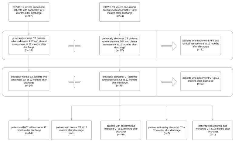
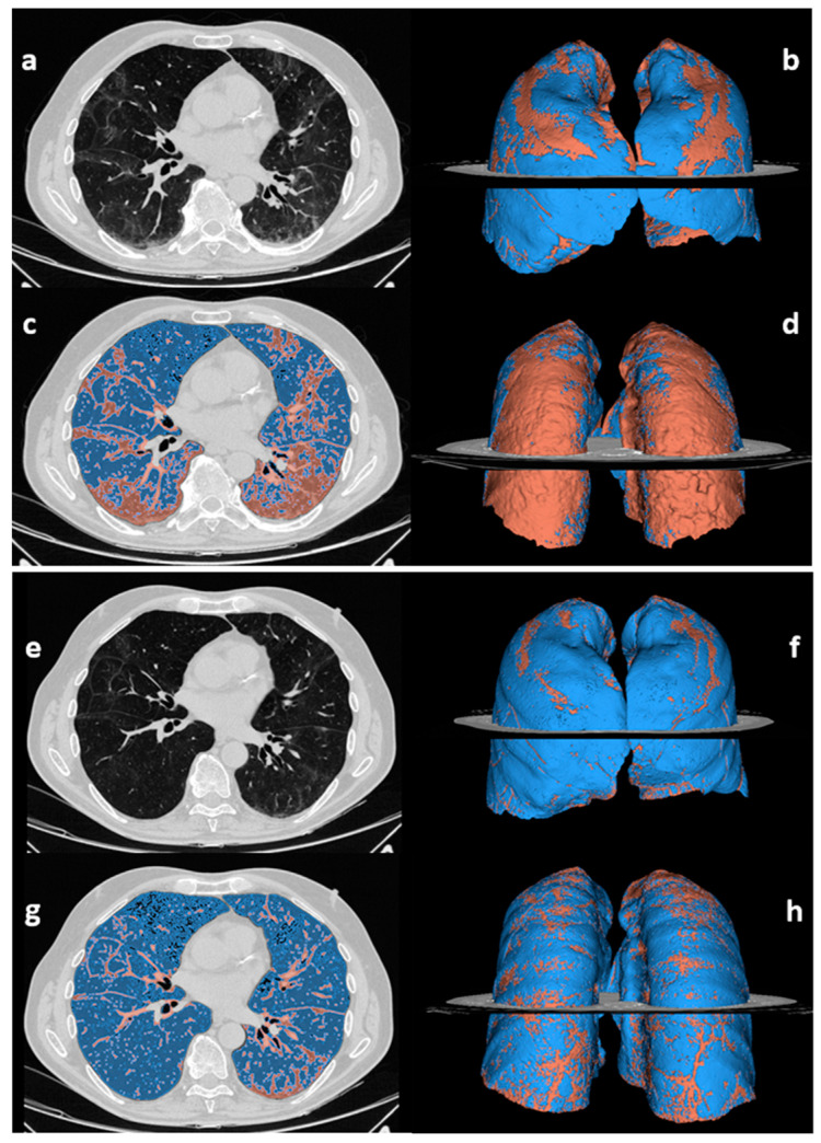
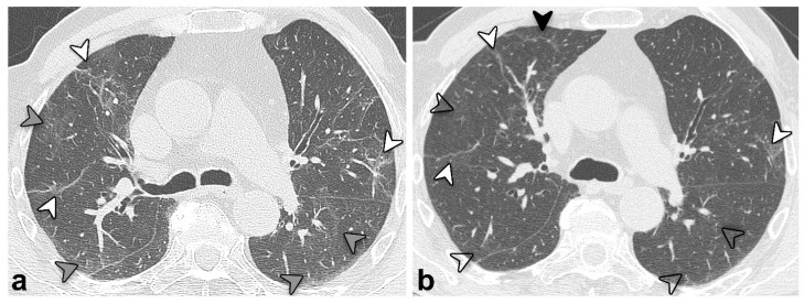
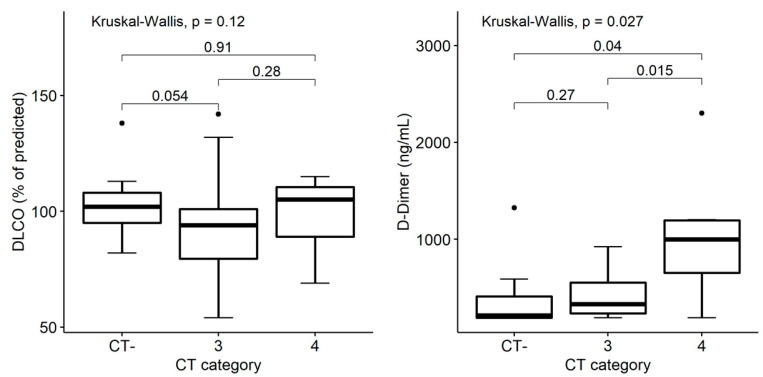
Long-term pulmonary sequelae in COVID-19 patients are currently under investigation worldwide. Potential relationships between blood sampling and functional and radiological findings are crucial to guide the follow-up. In this study, we collected and evaluated clinical status, namely symptoms and patients’ reported outcome, pulmonary function tests (PFT), laboratory tests, and radiological findings at 3- and 12-months post-discharge in patients admitted between 25 February and 2 May 2020, and who survived severe COVID-19 pneumonia. A history of chronic pulmonary disease or COVID-19-unrelated complications were used as exclusion criteria. Unenhanced CTs were analyzed quantitatively (compromising lung volume %) and qualitatively, with main patterns of: ground-glass opacity (GGO), consolidation, and reticular configuration. Patients were subsequently divided into groups based on their radiological trends and according to the evolution in the percentage of compromised lung volume. At 12 months post-discharge, seventy-one patients showed significantly improved laboratory tests and PFT. Among them, 63 patients also underwent CT examination: all patients with negative CT findings at three months (n = 14) had negative CT also at 12 months; among the 49/63 patients presenting CT alterations at three months, 1/49 (2%) normalized, 40/49 (82%) improved, 7/49 (14%) remained stably abnormal, and 1/49 (2%) worsened. D-dimer values were low in patients with normal CT and higher in cases with improved or stably abnormal CT (median values 213 vs. 329 vs. 1000 ng/mL, respectively). The overall compromised lung volume was reduced compared with three months post-discharge (12.3 vs. 14.4%, p < 0.001). In stably abnormal CT, the main pulmonary pattern changed, showing a reduction in GGO and an increase in reticular configuration. To summarize, PFT are normal in most COVID-19 survivors 12 months post-discharge, but CT structural abnormalities persist (although sensibly improved over time) and are associated with higher D-dimer values.
Keywords: COVID-19; lung diseases; respiratory function tests; severe acute respiratory syndrome coronavirus 2; tomography.
Conflict of interest statement
The Authors declare no conflict of interest.
Figures
References
-
- WHO COVID-19 Dashboard. Geneva: World Health Organization. 2020. [(accessed on 3 October 2022)]. Available online: https://covid19.who.int/
-
- Knight R., Walker V., Ip S., Cooper J.A., Bolton T., Keene S., Denholm R., Akbari A., Abbasizanjani H., Torabi F., et al. Association of COVID-19 With Major Arterial and Venous Thrombotic Diseases: A Population-Wide Cohort Study of 48 Million Adults in England and Wales. Circulation. 2022;146:892–906. doi: 10.1161/CIRCULATIONAHA.122.060785. - DOI - PMC - PubMed
-
- Tsagkaris C., Bilal M., Aktar I., Aboufandi Y., Tas A., Aborode A.T., Suvvari T.K., Ahmad S., Shkodina A., Phadke R., et al. Cytokine storm and neuropathological alterations in patients with neurological manifestations of COVID-19. Curr. Alzheimer Res. 2022 doi: 10.2174/1567205019666220908084559. - DOI - PubMed
-
- Baratella E., Roman-Pognuz E., Zerbato V., Minelli P., Cavallaro M.F.M., Cova M.A., Luzzati R., Lucangelo U., Sanson G., Friso F., et al. Potential links between COVID-19-associated pulmonary aspergillosis and bronchiectasis as detected by high resolution computed tomography. Front. Biosci. 2021;26:1607–1612. doi: 10.52586/5053. - DOI - PubMed
-
- Marando M., Fusi-Schmidhauser T., Tamburello A., Gauthier L.G., Rigamonti E., Argentieri G., Puligheddu C., Pagnamenta A., Valenti A., Pons M., et al. 1-year radiological, functional and quality-of-life outcomes in patients with SARS-CoV-2 pneumonia—A prospective observational study. NPJ Prim. Care Respir. Med. 2022;32:8. doi: 10.1038/s41533-022-00273-z. - DOI - PMC - PubMed
