Review
. 2022 Oct 21;8(5):2618-2638.
doi: 10.3390/tomography8050219.

Radiation Dose Reduction Opportunities in Vascular Imaging

Affiliations
Review

Radiation Dose Reduction Opportunities in Vascular Imaging

David Summerlin et al. Tomography. .

Abstract

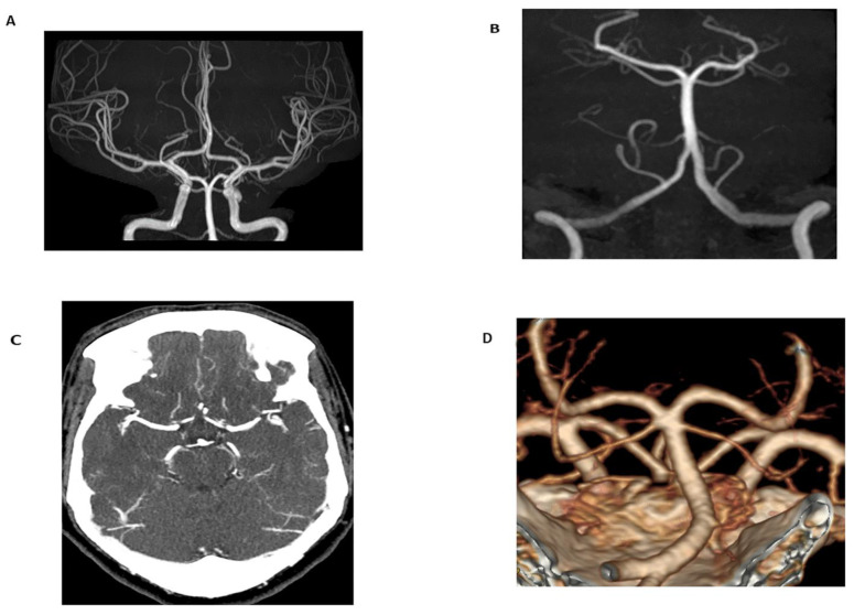
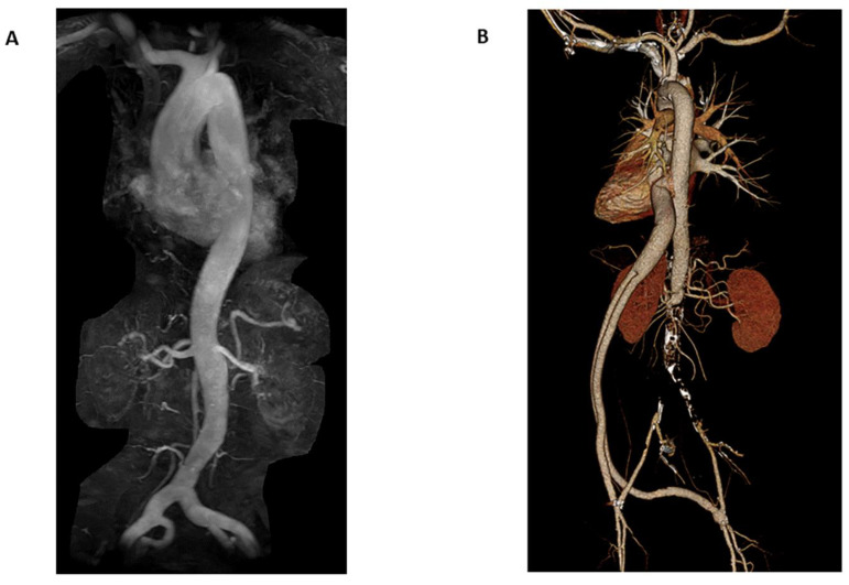
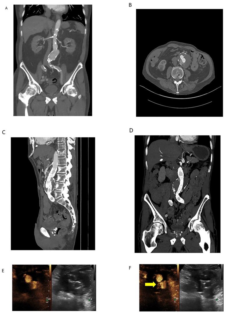
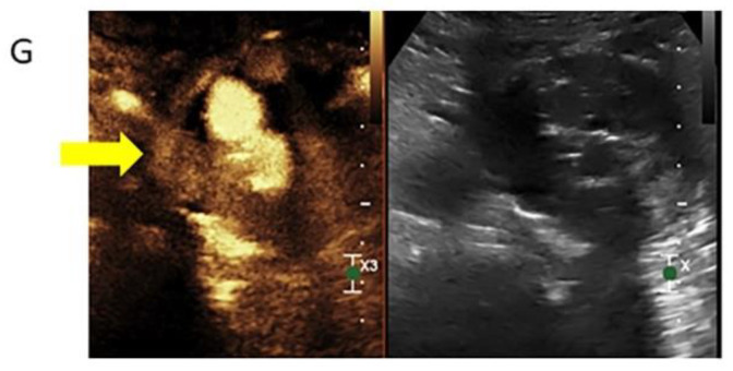
Computed tomography angiography (CTA) has been the gold standard imaging modality for vascular imaging due to a variety of factors, including the widespread availability of computed tomography (CT) scanners, the ease and speed of image acquisition, and the high sensitivity of CTA for vascular pathology. However, the radiation dose experienced by the patient during imaging has long been a concern of this image acquisition method. Advancements in CT image acquisition techniques in combination with advancements in non-ionizing radiation imaging techniques including magnetic resonance angiography (MRA) and contrast-enhanced ultrasound (CEUS) present growing opportunities to reduce total radiation dose to patients. This review provides an overview of advancements in imaging technology and acquisition techniques that are helping to minimize radiation dose associated with vascular imaging.

Keywords: computed tomography angiography; magnetic resonance angiography; radiation dose; radiation reduction.


Conflict of interest statement

The authors declare no conflict of interest.

Figures

Figure 1
Figure 1
Time-of-flight (TOF) maximum intensity projection (MIP) image of the Circle of Willis (A) and basilar artery (B) in a 24-year-old female obtained for migraines and dizziness. The magnetic resonance angiogram (MRA) demonstrated no flow-limiting stenosis or occlusion. Additionally, imaging was performed without radiation or intravenous contrast (technical specifications: FOV 200.00 mm, TR 25 ms, TE 3.5 ms, 3 T magnetic field strength). In comparison to TOF-MRA, axial CT angiogram (CTA) MIP images (C) and volume-rendered images (D) in a 60-year-old female obtained for evaluation of tinnitus demonstrated no flow-limiting stenosis or occlusion. 100 mL of Omnipaque 350 contrast was administered and the CTDIvol for the CTA of the head and neck was 59.6 mGy.
Figure 2
Figure 2
Coronal MIP MRA of the chest, abdomen, and pelvis (A) in a 66-year-old female for evaluation of possible aortic aneurysm demonstrates mild ectasia of the common iliac arteries bilaterally; however, no abdominal aortic aneurysm (AAA). 10 mL of Prohance contrast was administered. Coronal CTA volume rendered images (B) in a 70-year-old male with history of aortobifemoral bypass for suspected dissection demonstrates chronic occlusion of the infrarenal abdominal aorta and patent bypass. No dissection. 70 mL of Isovue contrast was administered with CTDIvol 19.2 mGy.
Figure 3
Figure 3
Coronal MIP image (A) demonstrating an infrarenal AAA in a 78-year-old male obtained prior to endovascular repair. Calcific atherosclerotic plaque and vessel patency well-demonstrated on this arterial phase CTA. 100 mL of Omnipaque 350 contrast was administered with CTDIvol of 7.68 mGy. Arterial phase source axial image (B) from a CTA demonstrating endovascular repair of the infrarenal AAA with persistent contrast flow into the excluded aneurysm sac, consistent with an endoleak. Sagittal (C) and coronal (D) MIP images are also shown. Lack of dynamic imaging limits classification of the type of endoleak; although, it was initially characterized as a type II endoleak by CTA. 100 mL of Isovue 370 contrast was administered with CTDIvol of 5.77 mGy. Subsequent contrast-enhanced ultrasound (CEUS, (E)) with dynamic contrast enhancement demonstrated a type III endoleak arising from the origin of the left iliac limb ((F), yellow arrow). The type III endoleak represents the major component and a large portion of the bolus empties into the aneurysm sac ((G), yellow arrow) with a small type II contribution arising from a right lumbar artery (not shown). Three boluses totaling 5 mL of Lumason were injected.
Figure 3
Figure 3
Coronal MIP image (A) demonstrating an infrarenal AAA in a 78-year-old male obtained prior to endovascular repair. Calcific atherosclerotic plaque and vessel patency well-demonstrated on this arterial phase CTA. 100 mL of Omnipaque 350 contrast was administered with CTDIvol of 7.68 mGy. Arterial phase source axial image (B) from a CTA demonstrating endovascular repair of the infrarenal AAA with persistent contrast flow into the excluded aneurysm sac, consistent with an endoleak. Sagittal (C) and coronal (D) MIP images are also shown. Lack of dynamic imaging limits classification of the type of endoleak; although, it was initially characterized as a type II endoleak by CTA. 100 mL of Isovue 370 contrast was administered with CTDIvol of 5.77 mGy. Subsequent contrast-enhanced ultrasound (CEUS, (E)) with dynamic contrast enhancement demonstrated a type III endoleak arising from the origin of the left iliac limb ((F), yellow arrow). The type III endoleak represents the major component and a large portion of the bolus empties into the aneurysm sac ((G), yellow arrow) with a small type II contribution arising from a right lumbar artery (not shown). Three boluses totaling 5 mL of Lumason were injected.

References

    1. Costello J.E., Cecava N.D., Tucker J.E., Bau J.L. CT Radiation Dose: Current Controversies and Dose Reduction Strategies. AJR Am. J. Roentgenol. 2013;201:1283–1290. doi: 10.2214/AJR.12.9720. - DOI - PubMed
    1. Albrecht M.H., Bickford M.W., Nance J.W., Zhang L., De Cecco C.N., Wichmann J.L., Vogl T.J., Schoepf U.J. State-of-the-Art Pulmonary CT Angiography for Acute Pulmonary Embolism. AJR Am. J. Roentgenol. 2017;208:495–504. doi: 10.2214/AJR.16.17202. - DOI - PubMed
    1. Baralo B., Samson P., Hoenig D., Smith A. Percutaneous kidney stone surgery and radiation exposure: A review. Asian J. Urol. 2019;7:10–17. doi: 10.1016/j.ajur.2019.03.007. - DOI - PMC - PubMed
    1. National Research Council, Division on Earth and Life Studies, Board on Radiation Effects Research, Committee to Assess Health Risks from Exposure to Low Levels of Ionizing Radiation . Health Risks from Exposure to Low Levels of Ionizing Radiation: BEIR VII Phase 2. National Academies Press; Washington, DC, USA: 2006.
    1. AAPM Position Statements, Policies and Procedures—Details 2018. [(accessed on 6 August 2022)]. Available online: https://www.aapm.org/org/policies/details.asp?type=PP&id=2548.

LinkOut - more resources