Review
. 2022 Oct 17;14(10):709.
doi: 10.3390/toxins14100709.

Mechanisms of Action of the Peptide Toxins Targeting Human and Rodent Acid-Sensing Ion Channels and Relevance to Their In Vivo Analgesic Effects

Affiliations
Review

Mechanisms of Action of the Peptide Toxins Targeting Human and Rodent Acid-Sensing Ion Channels and Relevance to Their In Vivo Analgesic Effects

Clément Verkest et al. Toxins (Basel). .

Abstract

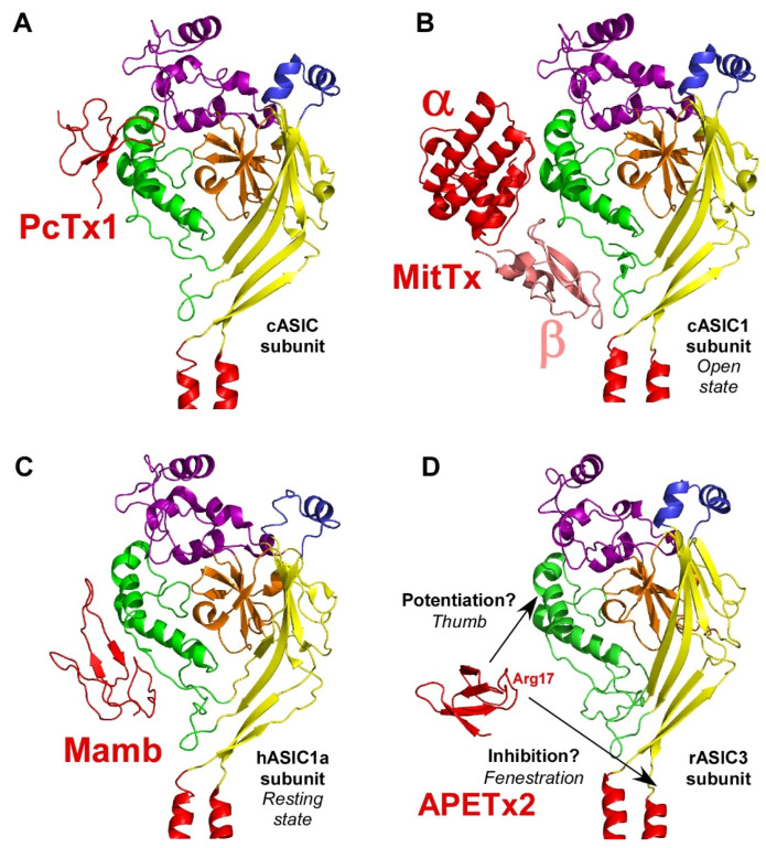
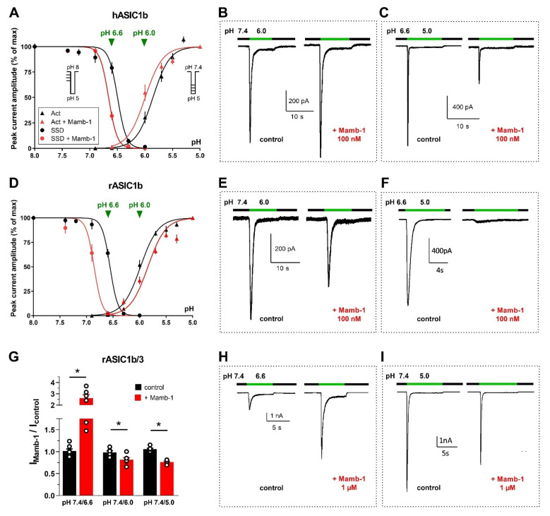
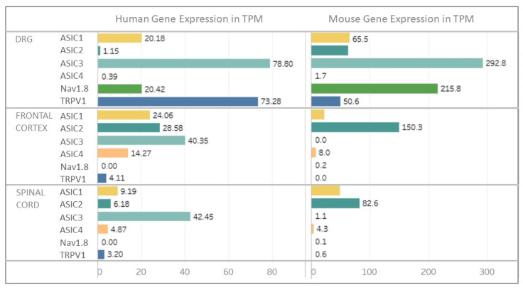
Acid-sensing ion channels (ASICs) are voltage-independent H+-gated cation channels largely expressed in the nervous system of rodents and humans. At least six isoforms (ASIC1a, 1b, 2a, 2b, 3 and 4) associate into homotrimers or heterotrimers to form functional channels with highly pH-dependent gating properties. This review provides an update on the pharmacological profiles of animal peptide toxins targeting ASICs, including PcTx1 from tarantula and related spider toxins, APETx2 and APETx-like peptides from sea anemone, and mambalgin from snake, as well as the dimeric protein snake toxin MitTx that have all been instrumental to understanding the structure and the pH-dependent gating of rodent and human cloned ASICs and to study the physiological and pathological roles of native ASICs in vitro and in vivo. ASICs are expressed all along the pain pathways and the pharmacological data clearly support a role for these channels in pain. ASIC-targeting peptide toxins interfere with ASIC gating by complex and pH-dependent mechanisms sometimes leading to opposite effects. However, these dual pH-dependent effects of ASIC-inhibiting toxins (PcTx1, mambalgin and APETx2) are fully compatible with, and even support, their analgesic effects in vivo, both in the central and the peripheral nervous system, as well as potential effects in humans.

Keywords: APETx2; ASIC; MitTx; PcTx1; mambalgin; nociception; pain; peptide; sodium channels; toxins.


Conflict of interest statement

The authors declare no conflict of interest.

Figures

Figure 1
Figure 1
Structure of ASICs. (A) Trimeric organization of ASICs (left panel: side view, right panel: top view). (B) Tridimensional skeletal model of a single subunit where variable regions between isoforms “a” and “b” of rat ASIC1 and ASIC2 are highlighted in gold. (C) Structure of a single subunit of chicken ASIC1 in resting state (the different sub-domains are shown in specific colors; PDB ID: 6vtl). (D) Skeletal 3D representation of a functional channel formed by the assembly of three subunits. A transparent grey surface was added to one subunit to delineate the interface between two adjacent subunits. Same colors as in (C) for the different sub-domains, and key structural domains mentioned on the right. Cytoplasmic N- and C-termini, whose structures are unknown, are not shown. Designed with PyMOL software.
Figure 2
Figure 2
Diversity of currents flowing through homo- and heterotrimeric cloned ASICs. Original current traces of rat heterologously expressed ASIC currents recorded from HEK293 cells depending on the composition in ASIC subunits, activated from pH 7.4 to the indicated test pH, at −60 mV. Homotrimeric channels result from the expression of only one type of ASIC subunit (indicated above each current), whereas heterotrimeric channels result from the co-expression of two different subunits (1:1 ratio in transfection). The corresponding current noted rASIC1b/1a, for example, results from the co-expression of rASIC1b and rASIC1a subunits.
Figure 3
Figure 3
pH-dependent gating mechanisms of ASICs and interaction with toxins. ASIC gating involves three conformational states. (A), high pH resting state, which is stabilized by the toxin mambalgin (see Section 2.4) (major domains involved are indicated with the same color code as in Figure 1). (B), low pH open state, which is stabilized by the toxin MitTx (see Section 2.3) and also partially by the toxin PcTx1 (see Section 2.2). (C), low pH desensitized state also promoted by PcTx1. To illustrate the recovery process in (A), the deprotonation mechanism of only one acidic pocket is presented. Blue (A), green (B) and red (C) arrows show critical conformational changes during recovery, activation and desensitization processes, respectively. For clarity, only two subunits are shown.
Figure 4
Figure 4
Toxins interfere with the pH-dependent gating of rodent and human ASICs. Schematic pH-dependent curves of normalized (I/Imax%) activation (Acti, green) and steady state desensitization (SSD, red) of heterologously expressed cloned rat and human ASIC1a and ASIC1b in the absence (solid line) and in the presence of PcTx1 (dashed line) (AD) and of rat and human ASIC1a in the presence and in the absence of mambalgin (Mamb) (E,F). All curves were adapted from published data. (A,B), rASIC1a (A) and hASIC1a (B) gating modulation by PcTx1. The toxin increases the apparent H+ affinity of rASIC1a current thus inducing a leftward shift of both the activation and SSD curves towards more alkaline pH values. (A), PcTx1 inhibitory effect on rASIC1a current from physiological conditioning pH 7.4 (black downward arrow) to every test pH is mostly due to its pH-dependent SSD promoting effect, whereas no more inhibition was observed from pH 8.0 (⊘) instead revealing a potentiation of the current at test pH in the activation curve pH range (7.2–6.2), due to the opposite potentiating effect by a leftward shift of the activation curve (black upward arrow) [29] (PcTx1 10 nM). (B), PcTx1 exerts almost no effect on the hASIC1a current maximally activated from conditioning pH 7.4 (⊘), an inhibitory effect on the hASIC1a current maximally activated from conditioning pH 7.2 (black downward arrow), and a potentiation on the hASIC1a current submaximally activated from conditioning pH 7.4 (black upward arrow). Curves adapted from [30,31] (PcTx1 1 nM), with the shift of activation curve deduced from the PcTx1-induced current potentiation at test pH 6.7 (green points, PcTx1 60 nM) [19]. (C,D), rASIC1b (C) and hASIC1b (D) gating modulation by PcTx1. The toxin promotes opening of rASIC1b and hASIC1b through a leftward shift of the activation curve towards less acidic pH with almost no effect on the SSD curve. Consequently, PcTx1 does no inhibit the current maximally activated from conditioning pH 7.4 (⊘), and potentiates the current submaximally activated from pH 7.4 to test pH 6.8–5.8 (black upward arrow). Curves adapted from [32] (PcTx1 100 nM), and the shift of hASIC1b activation curve is deduced from the PcTx1-induced potentiation of the current at test pH 6.3 (green points, PcTx1 60 nM) [19]. The effect of PcTx1 on hASIC1b SSD curve is not yet known. (E,F), rASIC1a (E) and hASIC1a (F) gating modulation by Mamb. Mamb inhibits rASIC1a and hASIC1a currents mainly by a rightward shift of the pH-dependent activation curve towards more acidic pH values. Curves for rASIC1a current adapted from [33] (Mamb-1, 200 nM), and for hASIC1a from [34] (Mamb-3, 10 nM; note that this concentration is below IC50 value of Mamb on hASIC1a (see Table 3) and that a higher shift could thus be expected with a higher Mamb concentration). Red and green arrows illustrate shifts (acidic rightward, alkaline leftward) in the pH-dependent curves of activation and/or SSD by toxins. Data on the gating modulation of hASIC1b and rASIC1b by mambalgin-1 are shown in another following figure.
Figure 5
Figure 5
Toxin binding sites on one ASIC subunit. (A) Structure of a single cASIC1 subunit (rotated view of the skeletal 3D representation shown in Figure 1C) in complex with PcTx1 (PDB ID: 3s3x) [51]. (B), Structure of a single cASIC1 subunit in complex with MitTx (heterodimeric complex of MitTx-α and MitTx-β (PDB ID: 4NTY) [60]. (C), Cryo-EM structure of a single hASIC1a subunit in complex with mambalgin-1 (Mamb) at pH 8.0 (PDB ID: 7CFT) [52]. (D), Model of a single rASIC3 subunit extrapolated from cASIC1 structure, along with APETx2 (PDB ID: 2MUB) at the same scale, with two potential binding sites (black arrows) [129]. Designed with PyMOL software.
Figure 6
Figure 6
Dual pH-dependent effects of mambalgin-1 on whole-cell currents flowing through hASIC1b (AC), rASIC1b (DF) and rASIC1b/3 (GI) heterologously expressed in HEK293 cells (unpublished data). (A) on hASIC1b current, mambalgin-1 (Mamb-1, 100 nM) induced a leftward shift of both the pH-dependent activation (Act) curve (pH0.5 shifted from 5.86 to 6.0, p = 0.02, Mann–Whitney test) supporting a potentiating effect, and of the pH-dependent SSD curve (pH0.5 shifted from 6.50 to 6.66, p = 0.004) supporting an inhibitory effect. The protocols used for activation and SSD are shown in inset in A (mean ± SEM; n = 4–12 cells per point). (B,C), original hASIC1b whole-cell current traces recorded at −60 mV illustrating the dual effects of Mamb-1 (100 nM, applied 30 s before the pH drop) on a current activated by a pH drop from 7.4 to 6.0 (potentiation, B), and on a current activated by a pH drop from 6.6 to 5.0 (inhibition, C). (D) on rASIC1b current, Mamb-1 (100 nM) induced a rightward shift of the pH-dependent activation curve (pH0.5 shifted from 5.98 to 5.85, p = 0.002) and a leftward shift of the pH-dependent SSD curve (pH0.5 shifted from 6.56 to 6.86, p = 0.0002), both supporting an inhibitory effect (same protocols and curve labels as in A; mean ± SEM; n = 4–12 cells per point). (E,F), original rASIC1b whole-cell current traces recorded in the same conditions as in B-C and illustrating the partial inhibition by Mamb-1 of a current activated by a pH drop from 7.4 to 6.0 (E), and the full inhibition of a current activated by a pH drop from 6.6 to 5.0 (F). (G) bar graph quantification with individual data points of the effects of Mamb-1 (1 µM) on rASIC1b/3 heteromeric current. Mamb-1 induced a potentiation of the current when activated by a pH drop from 7.4 to 6.6 (p = 0.02, Wilcoxon paired test), but a partial inhibition when activated by a pH drop from 7.4 to 6.0 (p = 0.03) or 5.0 (p = 0.03). Mean ± SEM; n = 6–7 cells per condition. * p < 0.05. (H,I), original rASIC1b/3 whole-cell current traces recorded in the same conditions as in (B,C) and illustrating the potentiating effect (H) and the partial inhibition (I) by Mamb-1 depending on the test pH value.
Figure 7
Figure 7
Expression of ASICs in rodent and human nervous tissues. Expression of ASIC genes in central and peripheral nervous system from RNAseq data for human and mouse ASIC1-4, Nav 1.8 and TRPV1 genes from dorsal root ganglia (DRG), frontal cortex and spinal cord. Nav 1.8 and TRPV1 channels were shown as typically expressed in nociceptor sensory neurons. X axis represents TPM: Transcripts Per Million. Adapted from website: https://sensoryomics.shinyapps.io/RNA-Data/ [175] (accessed on June 2022).

References

    1. Krishtal O.A., Pidoplichko V.I. A receptor for protons in the nerve cell membrane. Neuroscience. 1980;5:2325–2327. doi: 10.1016/0306-4522(80)90149-9. - DOI - PubMed
    1. Waldmann R., Bassilana F., de Weille J., Champigny G., Heurteaux C., Lazdunski M. Molecular cloning of a non-inactivating proton-gated Na+ channel specific for sensory neurons. J. Biol. Chem. 1997;272:20975–20978. doi: 10.1074/jbc.272.34.20975. - DOI - PubMed
    1. Waldmann R., Champigny G., Bassilana F., Heurteaux C., Lazdunski M. A proton-gated cation channel involved in acid-sensing. Nature. 1997;386:173–177. doi: 10.1038/386173a0. - DOI - PubMed
    1. Kellenberger S., Schild L., Ohlstein E.H. International Union of Basic and Clinical Pharmacology. XCI. Structure, Function, and Pharmacology of Acid-Sensing Ion Channels and the Epithelial Na+ Channel. Pharmacol. Rev. 2015;67:1–35. doi: 10.1124/pr.114.009225. - DOI - PubMed
    1. Rosenbaum T., Morales-Lazaro S.L., Islas L.D. TRP channels: A journey towards a molecular understanding of pain. Nat. Rev. Neurosci. 2022;23:596–610. doi: 10.1038/s41583-022-00611-7. - DOI - PubMed

Publication types

LinkOut - more resources