Review
. 2022 Oct 17;7(10):310.
doi: 10.3390/tropicalmed7100310.

Strongyloides stercoralis: A Neglected but Fatal Parasite

Affiliations
Review

Strongyloides stercoralis: A Neglected but Fatal Parasite

Viravarn Luvira et al. Trop Med Infect Dis. .

Abstract

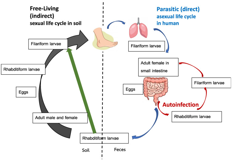
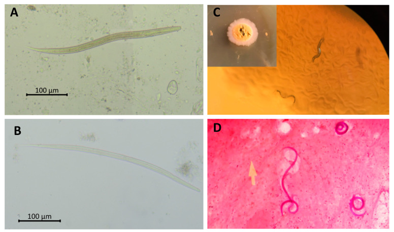
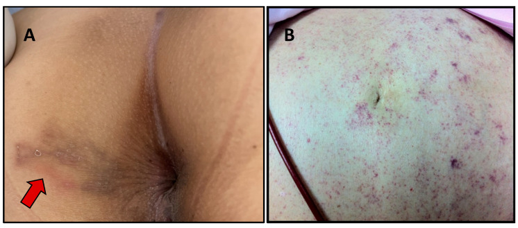
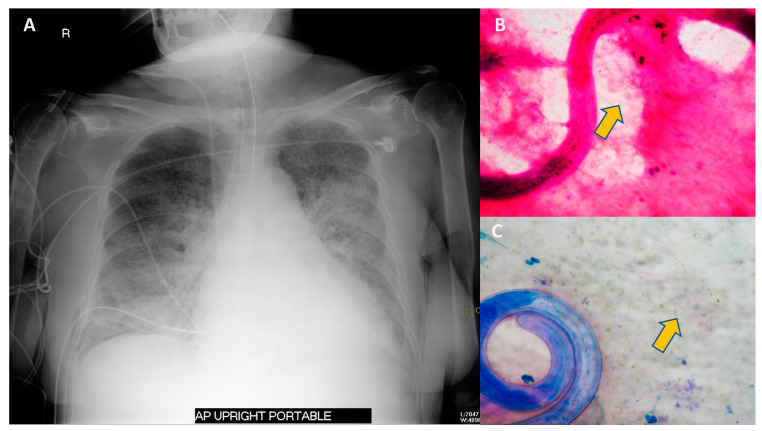
Strongyloidiasis is a disease caused by Strongyloides stercoralis and remains a neglected tropical infection despite significant public health concerns. Challenges in the management of strongyloidiasis arise from wide ranging clinical presentations, lack of practical high sensitivity diagnostic tests, and a fatal outcome in immunocompromised hosts. Migration, globalization, and increased administration of immunomodulators, particularly during the COVID-19 era, have amplified the global impact of strongyloidiasis. Here, we comprehensively review the diagnostic tests, clinical manifestations, and treatment of strongyloidiasis. The review additionally focuses on complicated strongyloidiasis in immunocompromised patients and critical screening strategies. Diagnosis of strongyloidiasis is challenging because of non-specific presentations and low parasite load. In contrast, treatment is simple: administration of single dosage ivermectin or moxidectin, a recent anthelmintic drug. Undiagnosed infections result in hyperinfection syndrome and disseminated disease when patients become immunocompromised. Thus, disease manifestation awareness among clinicians is crucial. Furthermore, active surveillance and advanced diagnostic tests are essential for fundamental management.

Keywords: Strongyloides stercoralis; disseminated strongyloidiasis; hyperinfection syndrome; immunocompromise; steroids; strongyloidiasis.


Conflict of interest statement

The authors declare no competing interest.

Figures

Figure 1
Figure 1
Life cycle of Strongyloides stercoralis. Adapted from reference no. [17].
Figure 2
Figure 2
Parasitological detection of Strongyloides stercoralis. Rhabditiform (A) and filariform; (B) larvae of S. stercoralis fresh smear. Migrating rhabditiform larvae in agar plate culture; (C). Gram staining of filariform larvae in sputum of a patient with S. stercoralis hyperinfection syndrome (100×) (D) Picture; (C) courtesy of Poom Adisakwattana.
Figure 3
Figure 3
Skin lesions that can be found in strongyloidiasis. Larva currens in the perianal area (arrow) (A) and periumbilical parasitic thumbprint purpura in a patient with disseminated strongyloidiasis (B). Picture (A) courtesy of Than Narkwiboonwong.
Figure 4
Figure 4
Chest radiography and sputum examination from a case of S. stercoralis hyperinfection syndrome in an immunocompromised patient. A 75-year-old Thai woman with temporal arteritis had been treated with prednisolone 20 mg/day for 4 months. She developed fever and diarrhea for 2 days prior to respiratory failure. The chest radiography revealed bilateral patchy infiltration (A). Sputum examination with Gram staining (B) and Modified Acid-Fast staining (C) revealed filariform larvae of S. stercoralis and a positive branching filamentous organism (arrow), indicating Nocardia species. Blood cultures grew Escherichia coli. She was diagnosed with S. stercoralis hyperinfection syndrome with concurrent Gram-negative bacteremia and pulmonary nocardiasis. She was treated with broad-spectrum cephalosporin, cotrimoxazole and ivermectin.

Similar articles

Cited by

References

    1. Schär F., Trostdorf U., Giardina F., Khieu V., Muth S., Marti H., Vounatsou P., Odermatt P. Strongyloides stercoralis: Global Distribution and Risk Factors. PLoS Negl. Trop. Dis. 2013;7:e2288. doi: 10.1371/journal.pntd.0002288. - DOI - PMC - PubMed
    1. Tamarozzi F., Martello E., Giorli G., Fittipaldo A., Staffolani S., Montresor A., Bisoffi Z., Buonfrate D. Morbidity Associated with Chronic Strongyloides stercoralis Infection: A Systematic Review and Meta-Analysis. Am. J. Trop. Med. Hyg. 2019;100:1305–1311. doi: 10.4269/ajtmh.18-0895. - DOI - PMC - PubMed
    1. Bisoffi Z., Buonfrate D., Montresor A., Requena-Méndez A., Muñoz J., Krolewiecki A.J., Gotuzzo E., Mena M.A., Chiodini P.L., Anselmi M., et al. Strongyloides stercoralis: A plea for action. PLoS Negl. Trop. Dis. 2013;7:e2214. doi: 10.1371/journal.pntd.0002214. - DOI - PMC - PubMed
    1. Keiser P.B., Nutman T.B. Strongyloides stercoralis in the Immunocompromised Population. Clin. Microbiol. Rev. 2004;17:208–217. doi: 10.1128/CMR.17.1.208-217.2004. - DOI - PMC - PubMed
    1. Caumes E., Keystone J.S. Acute strongyloidiasis: A rarity. Chronic strongyloidiasis: A time bomb! J. Travel Med. 2011;18:71–72. doi: 10.1111/j.1708-8305.2010.00492.x. - DOI - PubMed

LinkOut - more resources