. 2022 Nov;42(11):1333-1350.
doi: 10.1161/ATVBAHA.121.317749. Epub 2022 Oct 26.

Chronic E-Cigarette Use Impairs Endothelial Function on the Physiological and Cellular Levels

Affiliations

Chronic E-Cigarette Use Impairs Endothelial Function on the Physiological and Cellular Levels

Leila Mohammadi et al. Arterioscler Thromb Vasc Biol. 2022 Nov.

Erratum in

Abstract

Background: The harmful vascular effects of smoking are well established, but the effects of chronic use of electronic cigarettes (e-cigarettes) on endothelial function are less understood. We hypothesized that e-cigarette use causes changes in blood milieu that impair endothelial function.

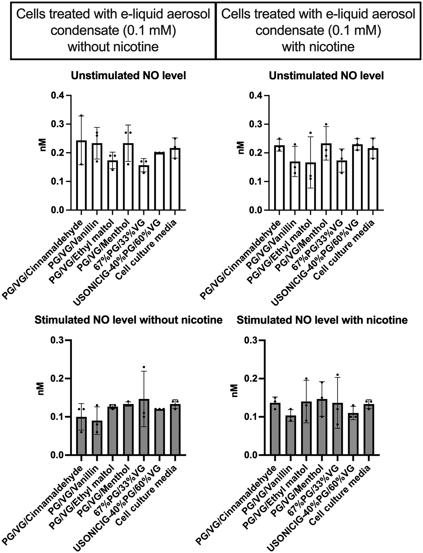
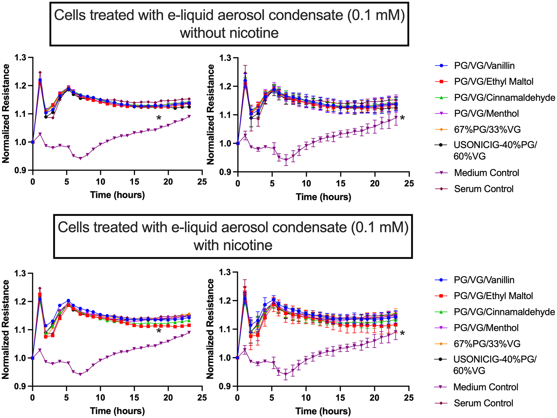
Methods: Endothelial function was measured in chronic e-cigarette users, chronic cigarette smokers, and nonusers. We measured effects of participants' sera, or e-cigarette aerosol condensate, on NO and H2O2 release and cell permeability in cultured endothelial cells (ECs).

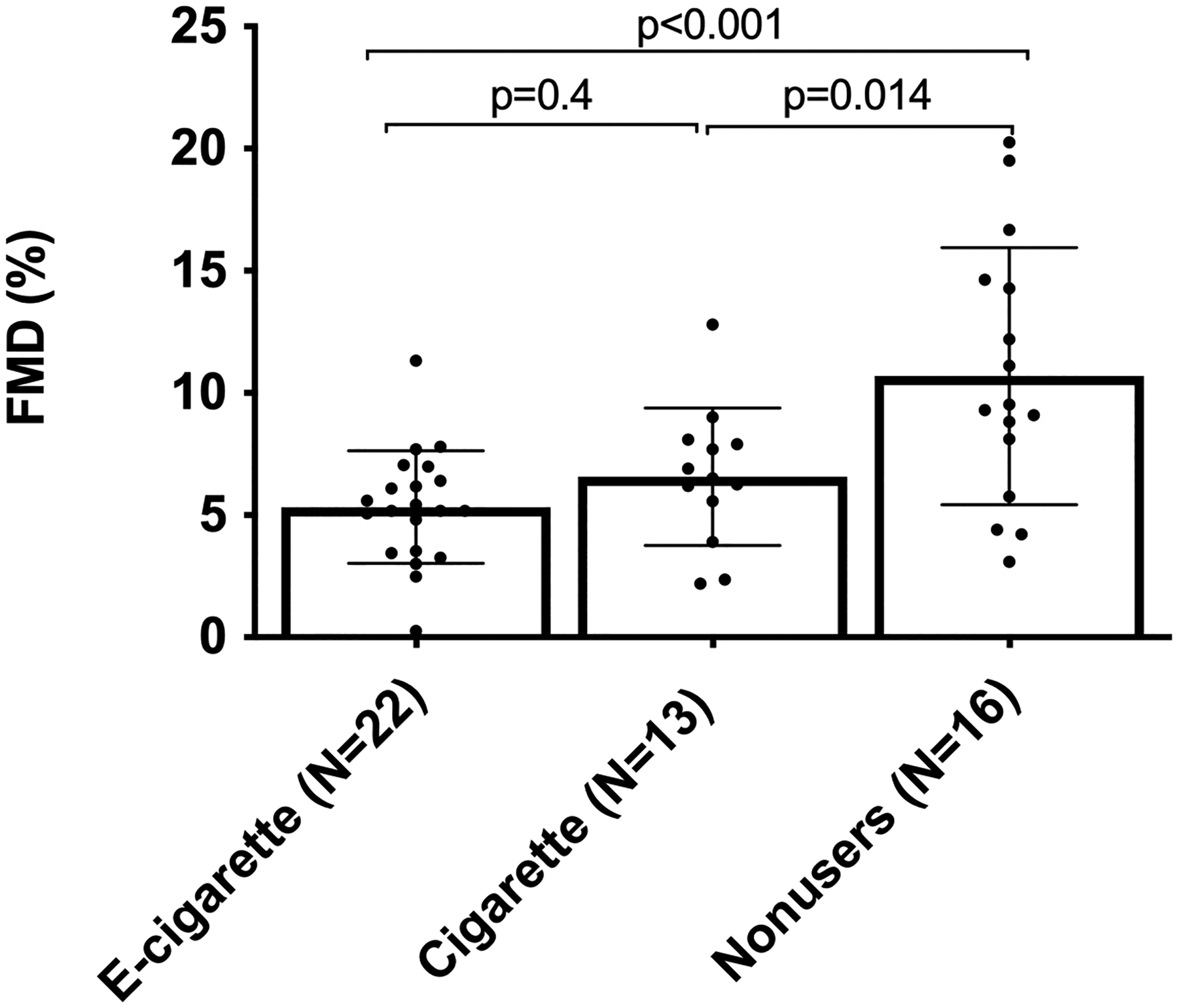
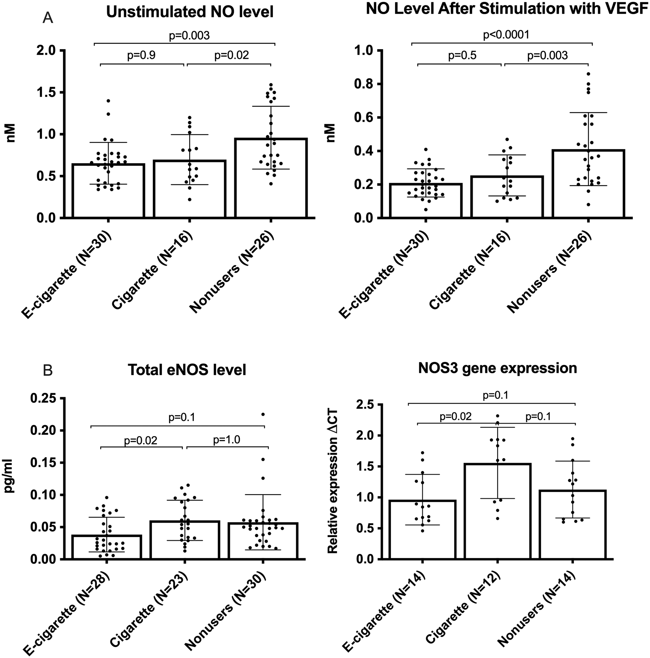
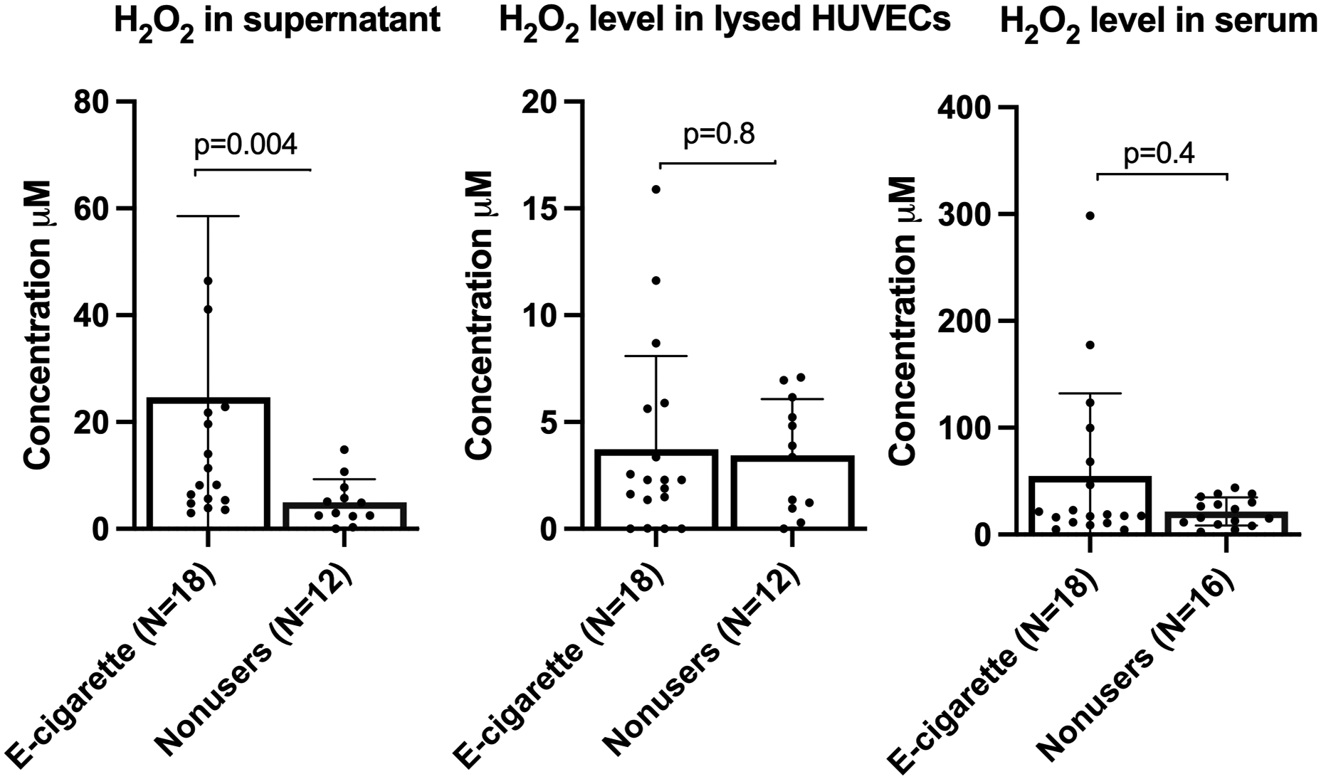
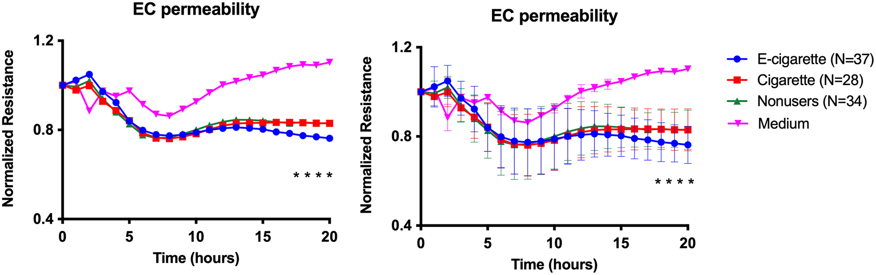
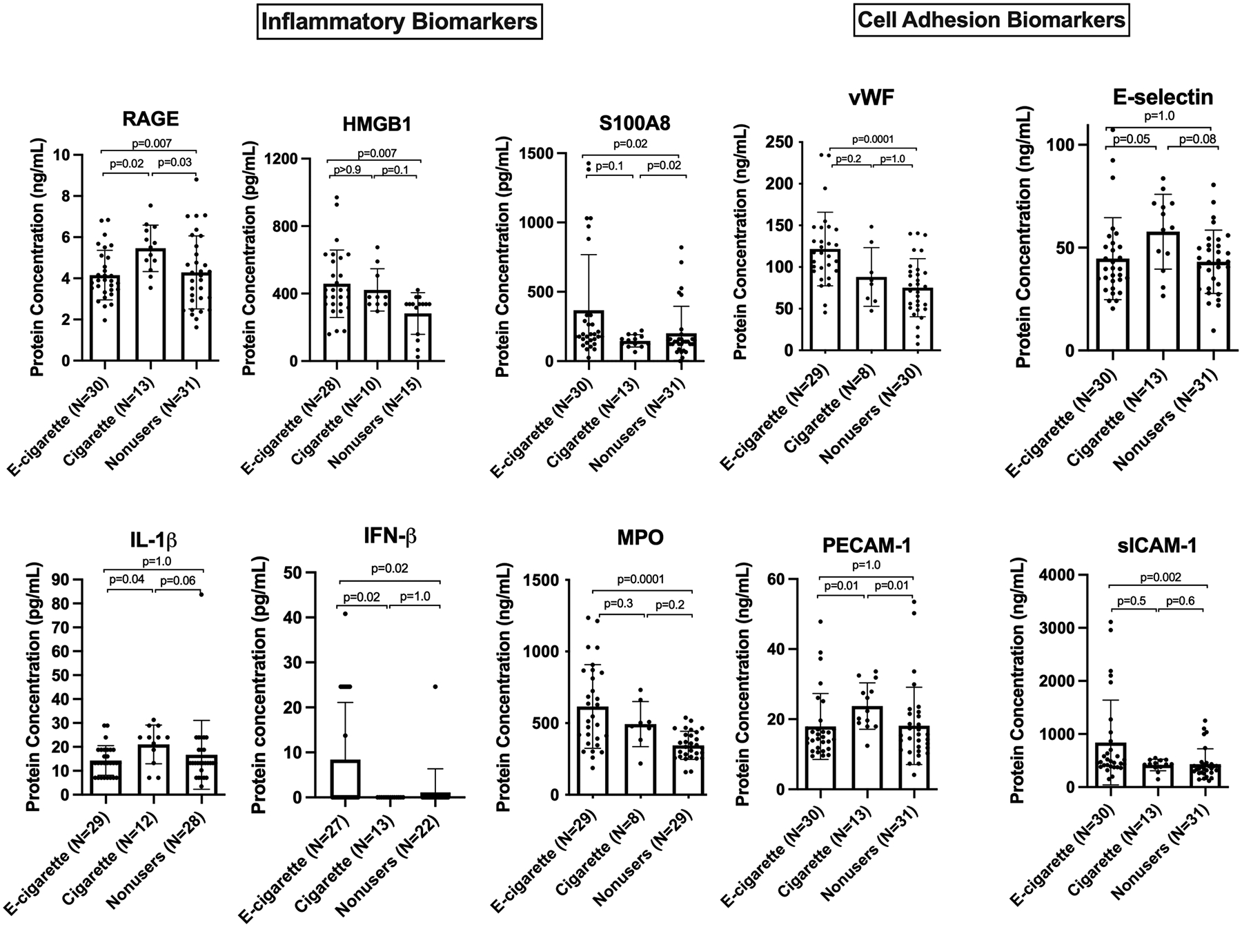
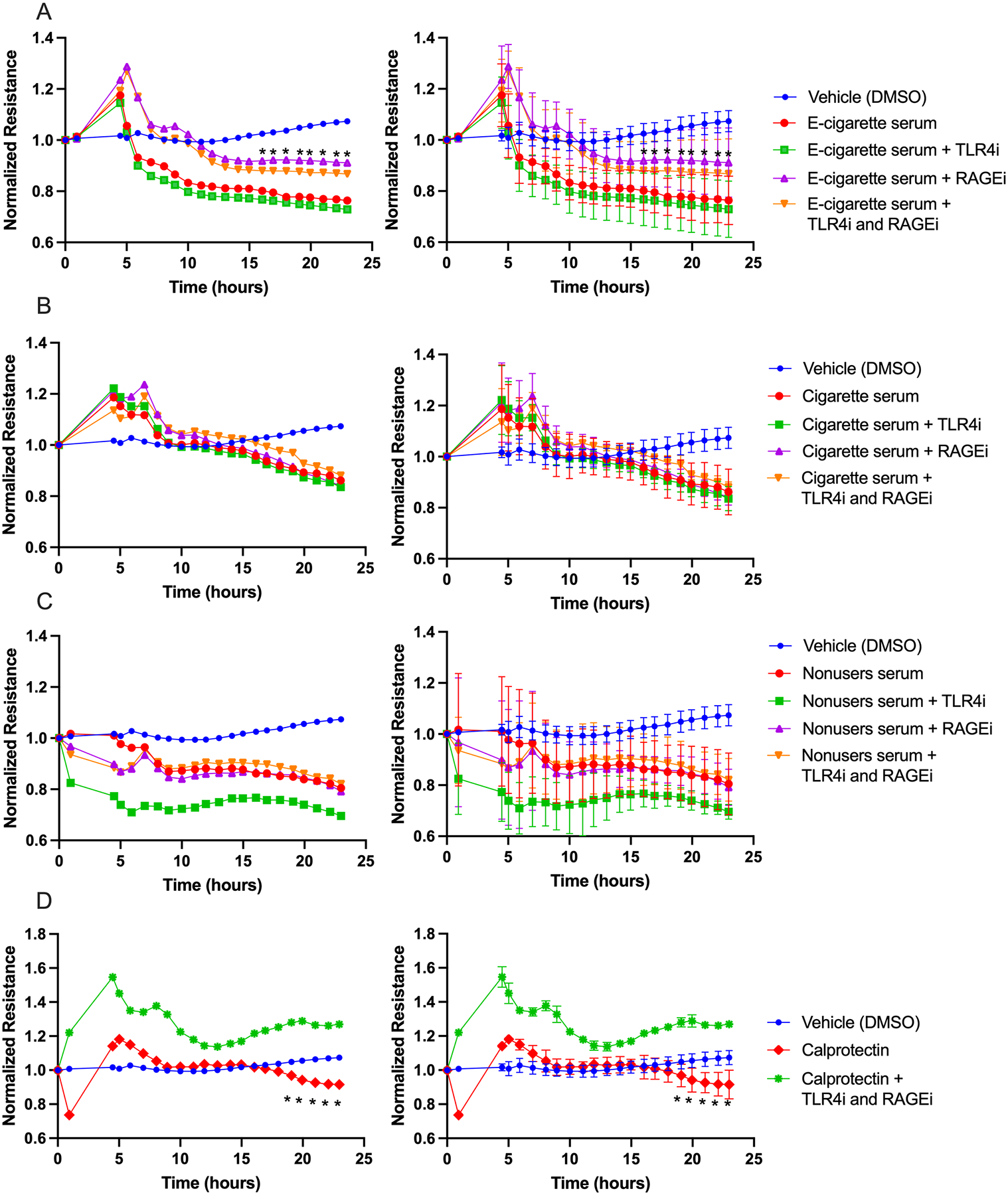
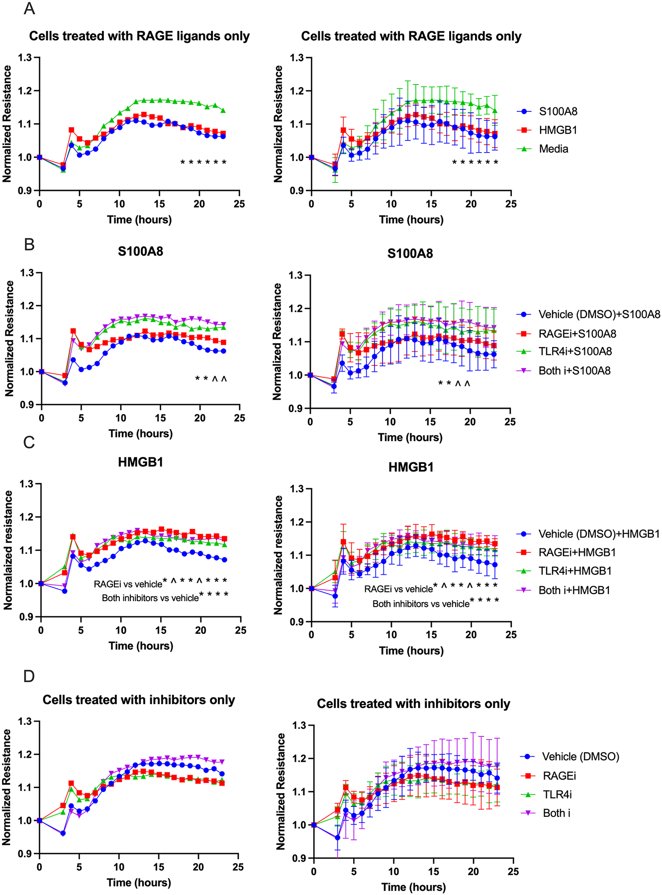
Results: E-cigarette users and smokers had lower flow-mediated dilation (FMD) than nonusers. Sera from e-cigarette users and smokers reduced VEGF (vascular endothelial growth factor)-induced NO secretion by ECs relative to nonuser sera, without significant reduction in endothelial NO synthase mRNA or protein levels. E-cigarette user sera caused increased endothelial release of H2O2, and more permeability than nonuser sera. E-cigarette users and smokers exhibited changes in circulating biomarkers of inflammation, thrombosis, and cell adhesion relative to nonusers, but with distinct profiles. E-cigarette user sera had higher concentrations of the receptor for advanced glycation end products (RAGE) ligands S100A8 and HMGB1 (high mobility group box 1) than smoker and nonuser sera, and receptor for advanced glycation end product inhibition reduced permeability induced by e-cigarette user sera but did not affect NO production.

Conclusions: Chronic vaping and smoking both impair FMD and cause changes in the blood that inhibit endothelial NO release. Vaping, but not smoking, causes changes in the blood that increase microvascular endothelial permeability and may have a vaping-specific effect on intracellular oxidative state. Our results suggest a role for RAGE in e-cigarette-induced changes in endothelial function.

Keywords: biomarker; cell adhesion; endothelial cell; inflammation; ligand.


Conflict of interest statement

Conflict of interest: None.

Figures

Figure 1.
Figure 1.. FMD of brachial artery.
Reduced brachial artery FMD in both e-cigarette users and cigarette smokers relative to nonusers. Group means were compared by one-way ANOVA with Holm-Šidák post-hoc adjustment. Bars=SD.
Figure 2.
Figure 2.. Effects of user sera (every participant) on NO production and eNOS gene expression.
(A) NO level released from cultured HUVECs before and after stimulation with VEGF. Stimulated values are lower than unstimulated values due to a 30-minute stimulated collection period vs. 12 hours for basal conditions. (B) eNOS protein and gene expression in HUVECs treated with individual serum samples. eNOS protein and gene expression were significantly lower in the e-cigarette group relative to smokers but not nonusers. There were 3 replicates per participant for NO and NOS3 measurements, and 2 replicates per participant for eNOS protein measurements. Group means were compared by one-way ANOVA with Holm-Šidák post-hoc adjustment. Bars=SD.
Figure 3.
Figure 3.. NO release from cells treated with e-liquid aerosol condensates at estimated physiological circulating level (0.1 mM).
Aerosol condensates did not significantly change NO production under basal or stimulated conditions (p<.05 required for significance; all p values were >0.3). Group means were compared by Kruskal-Wallis test with Dunn’s post-hoc adjustment. Each condition was run in triplicate. Bars=SD.
Figure 4.
Figure 4.. Significant increase in H2O2 level in cell culture supernatant exposed to e-cigarette users sera.
No significant changes in H2O2 level was observed in participants serum or lysed cells of all groups. Group means were compared by Mann-Whitney test. Bars=SD.
Figure 5.
Figure 5.. Increased microvascular endothelial permeability induced by e-cigarette user sera (every participant).
Graph shows resistance across a monolayer of HMVEC-Ls; lower resistance values correspond to greater permeability. *p<0.05 between e-cigarette user group compared to cigarette smoker and nonuser groups starting at 17 hours of incubation. There were 3 replicates per participant. Group means at each timepoint were compared by repeated measures ANOVA with Holm-Šidák post-hoc adjustment. Bars=SD; the graphs are equivalent but error bars have been omitted from one of them to enhance readability.
Figure 6.
Figure 6.. Endothelial cell permeability was not changed in cells treated with e-liquid aerosol condensate (0.1 mM).
Graphs show resistance across a monolayer of HMVEC-Ls; lower resistance values correspond to greater permeability. There were no significant differences in all comparisons except for Medium Control versus all other conditions (*p<0.05). Each condition was run in triplicate. Group means at each timepoint were compared by repeated measures ANOVA with Holm-Šidák post-hoc adjustment. Bars=SD; the left hand and right hand graphs are equivalent but error bars have been omitted from the left hand graphs to enhance readability.
Figure 7.
Figure 7.. Circulating biomarkers.
Chronic e-cigarette use led to changes in inflammatory and cell adhesion biomarkers relevant to endothelial dysfunction. Group means were compared by Kruskal-Wallis test with Dunn’s post-hoc adjustment. Bars=SD. (The mean level of IFN-β in cigarette smokers was below detection with one outlier with value >500 removed, and in nonusers, except for one person, it was below detection as well).
Figure 8.
Figure 8.. Inhibition of RAGE reduces the otherwise increased permeability from exposure to sera from e-cigarette users, but not from cigarette smokers or nonusers.
Graphs show resistance across a monolayer of HMVEC-Ls; lower resistance values correspond to greater permeability. (A) Exposure to e-cigarette user serum with and without inhibitors (*p<0.05 between e-cigarette serum and e-cigarette serum + RAGEi). (B) Similar exposure to cigarette smoker serum (no significant differences). (C) Similar exposure to nonuser serum (no significant differences). (D) With no serum, calprotectin led to increased cell permeability, which was reversed by adding both inhibitors (*p<0.05 between cells treated with only calprotectin and those treated with calprotectin and both inhibitors). For A-C, there were 10 participants per condition and 1 well per participant. For D, there were 3 replicates per condition. Group means at each timepoint were compared by repeated measures ANOVA with Holm-Šidák post-hoc adjustment. RAGEi = RAGE inhibitor, TLR4i = TLR4 inhibitor. Bars=SD; the left hand and right hand graphs are equivalent but error bars have been omitted from the left hand graphs to enhance readability.
Figure 9.
Figure 9.. Ligands of RAGE (S100A8 and HMGB1) increase cell permeability. RAGEi and TLR4i reduced cell permeability caused by the RAGE ligands.
Graphs show resistance across a monolayer of HMVEC-Ls; lower resistance values correspond to greater permeability. (A) Cells treated with each RAGE ligand alone had significantly greater permeability compared to the control starting at 17 hours of exposure (*p<.0.05 for media vs. S100A8 or HMBG1). (B) Cells treated with S100A8 followed by both inhibitors showed significant decrease in permeability starting at 19 hours of exposure compared to cells treated with each inhibitor or vehicle (*significant at p<.05 for both inhibitors vs. all other conditions; ^0.05<p<0.06 is also shown for reference). (C) Cells treated with HMGB1 followed by RAGEi showed a significant reduction in permeability compared to vehicle starting at 15 hours of exposure, and starting at 19 hours of exposure when treated with both inhibitors (see notes on graph; *significant at p<.05; ^0.05<p<0.06 is also shown for reference). (D) Inhibitors alone did not change cell permeability. Group means at each timepoint were compared by repeated measures ANOVA with Holm-Šidák post-hoc adjustment. RAGEi = RAGE inhibitor, TLR4i = TLR4 inhibitor. Bars=SD; the left hand and right hand graphs are equivalent but error bars have been omitted from the left hand graphs to enhance readability.

Comment in

References

    1. Fairchild AL, Bayer R and Lee JS. The E-Cigarette Debate: What Counts as Evidence? Am J Public Health. 2019;109:1000–1006. - PMC - PubMed
    1. Fetterman JL, Weisbrod RM, Feng B, Bastin R, Tuttle ST, Holbrook M, Baker G, Robertson RM, Conklin DJ, Bhatnagar A and Hamburg NM. Flavorings in Tobacco Products Induce Endothelial Cell Dysfunction. Arterioscler Thromb Vasc Biol. 2018;38:1607–1615. - PMC - PubMed
    1. Fetterman JL, Keith RJ, Palmisano JN, McGlasson KL, Weisbrod RM, Majid S, Bastin R, Stathos MM, Stokes AC, Robertson RM, Bhatnagar A and Hamburg NM. Alterations in Vascular Function Associated With the Use of Combustible and Electronic Cigarettes. J Am Heart Assoc. 2020;9:e014570. - PMC - PubMed
    1. Rao P, Liu J and Springer ML. JUUL and Combusted Cigarettes Comparably Impair Endothelial Function. Tob Regul Sci. 2020;6:30–37. - PMC - PubMed
    1. Buchanan ND, Grimmer JA, Tanwar V, Schwieterman N, Mohler PJ and Wold LE. Cardiovascular risk of electronic cigarettes: a review of preclinical and clinical studies. Cardiovasc Res. 2020;116:40–50. - PMC - PubMed

Publication types