Review
. 2022 Oct 28;8(43):eabn3298.
doi: 10.1126/sciadv.abn3298. Epub 2022 Oct 26.

Control of infection by LC3-associated phagocytosis, CASM, and detection of raised vacuolar pH by the V-ATPase-ATG16L1 axis

Affiliations
Review

Control of infection by LC3-associated phagocytosis, CASM, and detection of raised vacuolar pH by the V-ATPase-ATG16L1 axis

Yingxue Wang et al. Sci Adv. .

Abstract

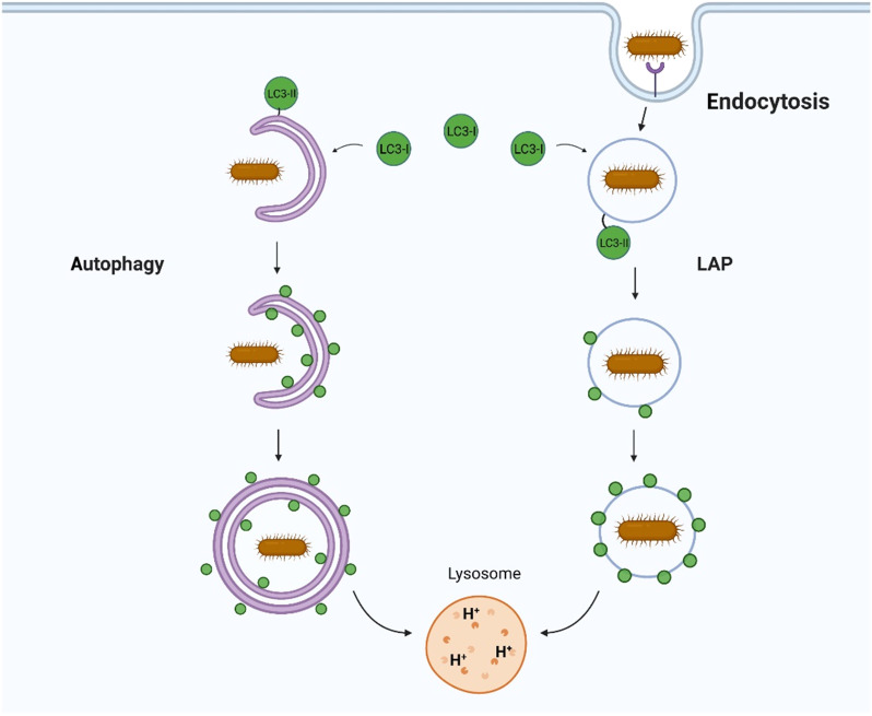
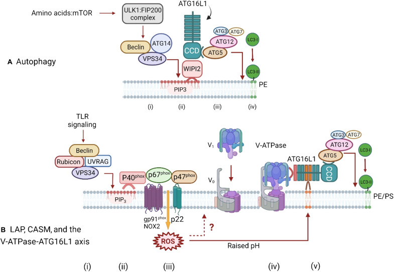
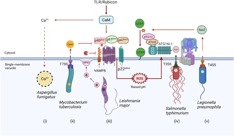
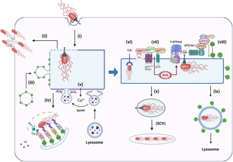
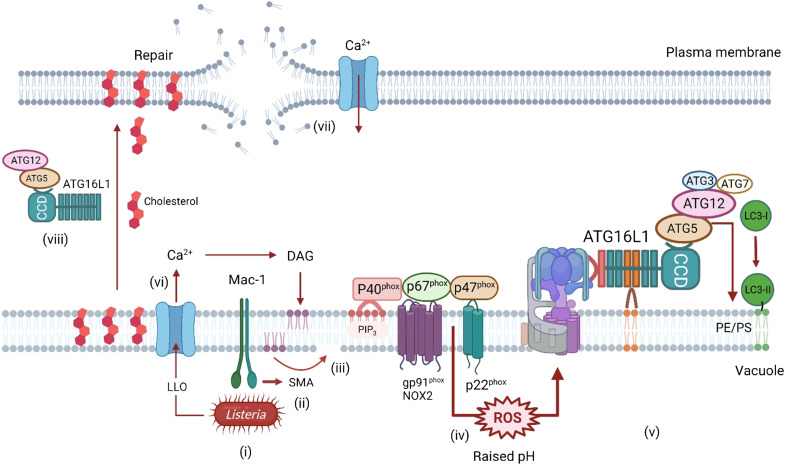
The delivery of pathogens to lysosomes for degradation provides an important defense against infection. Degradation is enhanced when LC3 is conjugated to endosomes and phagosomes containing pathogens to facilitate fusion with lysosomes. In phagocytic cells, TLR signaling and Rubicon activate LC3-associated phagocytosis (LAP) where stabilization of the NADPH oxidase leads to sustained ROS production and raised vacuolar pH. Raised pH triggers the assembly of the vacuolar ATPase on the vacuole membrane where it binds ATG16L1 to recruit the core LC3 conjugation complex (ATG16L1:ATG5-12). This V-ATPase-ATG16L1 axis is also activated in nonphagocytic cells to conjugate LC3 to endosomes containing extracellular microbes. Pathogens provide additional signals for recruitment of LC3 when they raise vacuolar pH with pore-forming toxins and proteins, phospholipases, or specialized secretion systems. Many microbes secrete virulence factors to inhibit ROS production and/or the V-ATPase-ATG16L1 axis to slow LC3 recruitment and avoid degradation in lysosomes.


Figures

Fig. 1.
Fig. 1.. Autophagy and LAP provide different ways to control infection.
Canonical autophagy conjugates LC3 to double-membrane autophagosomes that engulf pathogens in the cytosol. LAP conjugates LC3 to endosomes and phagosomes containing extracellular pathogens as they enter cells. In both cases, conjugation of LC3 to membranes containing pathogens facilitates fusion with lysosomes, leading to pathogen degradation.
Fig. 2.
Fig. 2.. Pathways for ATG16L1-mediated conjugation of LC3 to membranes during autophagy and LAP/CASM conjugation via the V-ATPase-ATG16L1 axis.
(A) Autophagy. Autophagy is activated in response to a fall in amino acids, which leads to inhibition of mammalian target of rapamycin (mTOR) and activation of the ULK1/FIP200 initiation complex (i) and downstream activation of the PI3K complex containing Beclin1, ATG14, and VPS34. The PI3K activity of VPS34 generates PIP3 in autophagosome membranes, which provide a platform for binding of WIPI2 (ii). WIPI2 binds to the coiled coil domain (CCD) of ATG16L1, leading to recruitment of the LC3 conjugation complex (ATG5-ATG12, ATG3, and ATG7). The conjugation reaction results in conversion of LC3I to LC3II and covalent binding of LC3 to PE in the autophagosome membrane (iii and iv). (B) LAP/CASM and the V-ATPase-ATG16L1 axis. LC3 conjugation in phagocytic cells is activated by TLR signaling through a complex containing Rubicon, Beclin1, UVRAG, VPS15, and VPS34 (i). TLR signaling activates VPS34 within the complex leading to generation of PIP3 in phagosome membranes (ii) to generate a binding site for p40phox that stabilizes the NADPH complex (NOX2, p47phox, p40phox, and p67phox). At the same time, binding of Rubicon to p22phox increases production of reactive oxygen species (ROS). Generation of ROS increases the pH in the lumen of the phagosome (iii) stimulating assembly of the Vo V1 subunits of Vo-V1 (iv). It is also possible that ROS can act directly on V-ATPase (?). V-ATPase binds to the WD domain of ATG16L1, leading to recruitment of the LC3 conjugation complex (ATG5-ATG12, ATG3, and ATG7) to the phagosome and conjugation of LC3 to phosphatidylserine (PS) and phosphatidylethanolamine (PE) in the phagosome membrane (v). A similar ROS-dependent pathway involving assembly of V-ATPase operates in nonphagocytic cells, but the precise components of the NADPH oxidase complex are unclear.
Fig. 3.
Fig. 3.. Recruitment of LC3 to membranes during STING signaling.
Microbial DNA in the cytoplasm activates cGAS (i). The cyclic GM-AMP synthase activity of cGAS generates cyclic GMP-AMP (cGAMP) (ii). Binding of cGAMP to STING releases STING from the ER, allowing transport in COPII-coated vesicles to Golgi membranes (iii). STING facilitates assembly of the Vo-V1 subunits of V-ATPase (iv). V-ATPase binds to the WD domain of ATG16L1, leading to recruitment of the LC3 conjugation complex (ATG5-ATG12, ATG3, and ATG7) and conjugation of LC3 to PE in the vesicle membrane. The precise identity of the vesicle membranes remains unclear, but they could be derived from the Golgi or endosomes.
Fig. 4.
Fig. 4.. The SNX5-ATG14 axis and viral infection.
The diagram uses influenza virus to illustrate responses to a broad range of viruses. Uptake of viruses into endosomes generates a signal (i) that recruits SNX5. SNX5 binds to ATG14 within the PI3K complex (VPS15, VPS34, Beclin1, and ATG14). VPS34 generates PIP3 in endosome membranes (ii), providing a binding site for WIPI2. The C-terminal BAR domain of SNX5 may sense (or generate) curvature in endosome membranes to enhance recruitment of ATG14 and generation of PIP3. WIPI2 binds to the CCD of ATG16L1, leading to recruitment of the LC3 conjugation complex (ATG5-ATG12, ATG3, and ATG7) (iii) and covalent binding of LC3 to PE and/or PS in the endosome membrane (iv). The V-ATPase-ATG16L1 axis may be activated when viruses generate pores in endosomes to facilitate delivery of genomes into the cytosol. This may involve fusion of capsids or envelopes with the endosome membrane (v) and/or synthesis of pore-forming proteins (viroporins). In the diagram, the M2 proton channel of influenza virus raises the pH of endosomes when inserted into the endosome membrane (vi). Raised pH leads to assembly of V-ATPase (vii) and recruitment of ATG16L1 and conjugation of LC3 to PE and/or PS (viii).
Fig. 5.
Fig. 5.. Overview of interactions between pathogens and LAP/CASM.
Pathogen entry activates TLR signaling followed by Rubicon-mediated assembly of the p67phox:p40phox:p47phox NADPH/NOX2 (gp91phox, p22phox) complex on the vacuole. Production of ROS raises vacuolar pH to activate the V-ATPase-ATG16L1 axis to conjugate LC3 to vacuolar membranes. The V-ATPase-ATG16L1 axis can also be activated when vacuolar membranes are damaged during assembly of membrane pores and translocons by pathogens. Several pathogens inhibit this pathway to slow delivery to lysosomes. Melanin in the spores of Aspergillus sequesters Ca++ required for recruitment of calmodulin (CaM) to the phagosome membrane. (i) CaM facilitates assembly of the p91phox:p40phox:p47phox complex. Mycobacterium use a type 7 secretion system (T7SS) to generate a 5-nm pore to deliver CpsA into the cytosol (ii). CpsA binds p40phox, and p47phox inhibits ROS production. Leishmania secrete exosomes containing GP63 (iii), which fuse with the phagosome membrane to deliver GP63 into the cytosol. GP63 is a metalloprotease that degrades VAMP to slow assembly of the p67phox:p40phox:p47phox complex. Salmonella assemble a type 3 secretion system (TSS3) to deliver SopF into the cytosol (iv). SopF inhibits binding of the WD domain of ATG16L1 to V-ATPase to inhibit the V-ATPase-ATG16L1 axis and conjugation of LC3. Legionella assemble a type 4 secretion system (T4SS) to deliver RavZ into the cytosol (v). RavZ is a protease that releases LC3 from the vacuole by deconjugating LC3 from PE.
Fig. 6.
Fig. 6.. Recruitment of LC3 to membranes during Salmonella infection.
Effectors encoded by SPI-1 are injected across the plasma membrane and stimulate actin rearrangements to facilitate invasion of the cell (i). A small number (10 to 20%) of bacteria escape to the cytosol and can hyperreplicate, causing cell death (ii). Assembly of the T3SS translocon generates pores in the endosome that can induce recruitment of LC3 and autophagosomes (iii) to stabilize the endosome and slow release of Salmonella into the cytosol, and at the same time, damaged endosomes and exposed bacteria can be ubiquitinated and recognized by cytosolic autophagy cargo sensors such as galectin 8 (Gal-8), NDP52, optineurin (OPN), and p62 and delivered to lysosomes for degradation (iv). Release of Ca2+ into the cytosol can activate synaptotagmin VII (SynVII)–mediated lysosome repair (v). Salmonella retained in early endosomes trigger TLR signaling (vi) to activate NADPH oxidase to generate ROS, leading to assembly of V-ATPase (vii). Pores generated by the T3SS translocon can also induce recruitment of V-ATPase to vacuoles containing Salmonella. V-ATPase binds the WD domain of ATG16L1, leading to recruitment of the LC3 conjugation complex (ATG5-ATG12, ATG3, and ATG7) and conjugation of LC3 to PE in the vacuole membrane (viii) and transport to lysosomes (ix). Binding of the WD domain to V-ATPase is inhibited by virulence factor SopF. If vacuoles containing salmonella are repaired, they maintain low pH, allowing effectors encoded by SPI-2 to modify the endosome to produce salmonella-containing vacuoles (SCVs), which provide a niche for slow replication in membrane compartments separated from autophagy and other innate sensors of infection (x).
Fig. 7.
Fig. 7.. Recruitment of LC3 to membranes during Listeria infection.
Binding of Listeria to Mac-1 in vacuoles in macrophages (i) activates sphingomyelinase (SMA), which removes the phosphocholine head group from sphingomyelin to generate ceramide (ii and iii). Ceramide-enriched microdomains facilitate assembly of the NADPH oxidase/NOX2 complex and enhance production of ROS (iv), which leads to assembly of V-ATPase and recruitment of ATG16L1:ATG5-ATG12 to conjugate LC3 to the vacuole membrane (v). LLO is a pore-forming toxin release by Listeria. Damage to the vacuole and subsequent entry of Ca2+ into the cytosol (vi) induce production of DAG, which activates the NADPH oxidase/NOX2 complex directly. Damage to the plasma membrane by LLO and entry of Ca2+ into the cytosol trigger a membrane repair pathway, which is dependent on the WD domain of ATG16L1 and ATG5-ATG12 (vii and viii). This may involve transport of cholesterol from vacuoles to the plasma membrane and lysosome-mediated membrane repair.

References

    1. Mizushima N., A brief history of autophagy from cell biology to physiology and disease. Nat. Cell Biol. 20, 521–527 (2018). - PubMed
    1. Xu Y., Jagannath C., Liu X.-D., Sharafkhaneh A., Kolodziejska K. E., Eissa N. T., Toll-like receptor 4 is a sensor for autophagy associated with innate immunity. Immunity 27, 135–144 (2007). - PMC - PubMed
    1. Sanjuan M. A., Dillon C. P., Tait S. W. G., Moshiach S., Dorsey F., Connell S., Komatsu M., Tanaka K., Cleveland J. L., Withoff S., Green D. R., Toll-like receptor signalling in macrophages links the autophagy pathway to phagocytosis. Nature 450, 1253–1257 (2007). - PubMed
    1. Huang J., Canadien V., Lam G. Y., Steinberg B. E., Dinauer M. C., Magalhaes M. A., Glogauer M., Grinstein S., Brumell J. H., Activation of antibacterial autophagy by NADPH oxidases. Proc. Natl. Acad. Sci. U.S.A. 106, 6226–6231 (2009). - PMC - PubMed
    1. Florey O., Gammoh N., Kim S. E., Jiang X., Overholtzer M., V-ATPase and osmotic imbalances activate endolysosomal LC3 lipidation. Autophagy 11, 88–99 (2015). - PMC - PubMed