Systematic lung ultrasound in Omicron-type vs. wild-type COVID-19
- PMID: 36288539
- PMCID: PMC9620376
- DOI: 10.1093/ehjci/jeac212
Systematic lung ultrasound in Omicron-type vs. wild-type COVID-19
Abstract
Aims: Preliminary data suggested that patients with Omicron-type-Coronavirus-disease-2019 (COVID-19) have less severe lung disease compared with the wild-type-variant. We aimed to compare lung ultrasound (LUS) parameters in Omicron vs. wild-type COVID-19 and evaluate their prognostic implications.
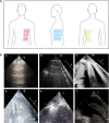
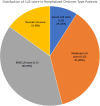
Methods and results: One hundred and sixty-two consecutive patients with Omicron-type-COVID-19 underwent LUS within 48 h of admission and were compared with propensity-matched wild-type patients (148 pairs). In the Omicron patients median, first and third quartiles of the LUS-score was 5 [2-12], and only 9% had normal LUS. The majority had either mild (≤5; 37%) or moderate (6-15; 39%), and 15% (≥15) had severe LUS-score. Thirty-six percent of patients had patchy pleural thickening (PPT). Factors associated with LUS-score in the Omicron patients included ischaemic-heart-disease, heart failure, renal-dysfunction, and C-reactive protein. Elevated left-filling pressure or right-sided pressures were associated with the LUS-score. Lung ultrasound-score was associated with mortality [odds ratio (OR): 1.09, 95% confidence interval (CI): 1.01-1.18; P = 0.03] and with the combined endpoint of mortality and respiratory failure (OR: 1.14, 95% CI: 1.07-1.22; P < 0.0001). Patients with the wild-type variant had worse LUS characteristics than the matched Omicron-type patients (PPT: 90 vs. 34%; P < 0.0001 and LUS-score: 8 [5, 12] vs. 5 [2, 10], P = 0.004), irrespective of disease severity. When matched only to the 31 non-vaccinated Omicron patients, these differences were attenuated.
Conclusion: Lung ultrasound-score is abnormal in the majority of hospitalized Omicron-type patients. Patchy pleural thickening is less common than in matched wild-type patients, but the difference is diminished in the non-vaccinated Omicron patients. Nevertheless, even in this milder form of the disease, the LUS-score is associated with poor in-hospital outcomes.
Keywords: COVID-19; clinical outcomes; lung ultrasound; risk stratification.
© The Author(s) 2022. Published by Oxford University Press on behalf of the European Society of Cardiology. All rights reserved. For permissions, please email: journals.permissions@oup.com.
Conflict of interest statement
Conflict of interest: None declared.
Figures


References
-
- Bouhemad B, Mongodi S, Via G, Rouquette I. Ultrasound for “lung monitoring” of ventilated patients. Anesthesiology 2015;122:437–47. - PubMed
MeSH terms
LinkOut - more resources
Full Text Sources
Medical
Research Materials

