Review
. 2022 Nov 2;110(21):3484-3496.
doi: 10.1016/j.neuron.2022.10.006. Epub 2022 Oct 7.

The neurobiology of long COVID

Affiliations
Review

The neurobiology of long COVID

Michelle Monje et al. Neuron. .

Abstract

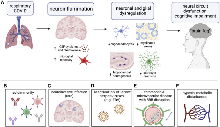
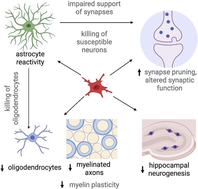
Persistent neurological and neuropsychiatric symptoms affect a substantial fraction of people after COVID-19 and represent a major component of the post-acute COVID-19 syndrome, also known as long COVID. Here, we review what is understood about the pathobiology of post-acute COVID-19 impact on the CNS and discuss possible neurobiological underpinnings of the cognitive symptoms affecting COVID-19 survivors. We propose the chief mechanisms that may contribute to this emerging neurological health crisis.

Keywords: COVID-19; EBV; HSV; PAISs; SARS-CoV-2; astrocytes; autoimmunity; blood-brain-barrier; cognitive impairment; hippocampal neurogenesis; long COVID; microglia; microvascular disease; myelin; post-acute infection syndromes.


Conflict of interest statement

Declaration of interests M.M. holds equity in MapLight Therapeutics and Syncopation Life Sciences. A.I. holds equity in RIGImmune and Xanadu Bio. M.M. is on the advisory board for Neuron.

Figures

Figure 1
Figure 1
Possible mechanisms contributing to COVID-19-related cognitive impairment (A) Respiratory system inflammation causes inflammation of the nervous system through systemic chemokines and other possible mechanisms. CNS cytokines, chemokines, and reactive microglia dysregulate multiple neural cell types, disrupt myelin homeostasis and plasticity, impair hippocampal neurogenesis, and induce neurotoxic astrocyte reactivity, each of which can impair neural circuit function and thus cognition. (B) Anti-neural autoantibodies and T cells can cause autoimmune encephalitis in patients with COVID-19 and could contribute to ongoing immune-mediated injury. (C) Neuroinvasive infection is rarely detected but can occur. (D) COVID-19 can trigger reactivation of latent herpesvirus infections, most prominently EBV, which can in turn incite further inflammation. (E) Neurovascular dysfunction, including blood-brain-barrier disruption (astrocyte endfeet, green) with consequent leakage of fibrinogen (blue linear molecule represented both intravascularly and in the extravascular space) and other pro-inflammatory molecules, and thrombosis (platelets, tan) can contribute to neural inflammation and injury. (F) In severe COVID-19, hypoxia and other metabolic disturbances from pulmonary and multi-organ dysfunction can cause nervous system injury. Figure created with BioRender.
Figure 2
Figure 2
Demonstrated (black text) and hypothesized (gray text) downstream consequences of reactive microglia in neuro-COVID Reactive microglia (red) can induce complex cellular dysregulation affecting neurons and oligodendrocytes. Demonstrated effects of reactive microglia after COVID-19 include a reduction in oligodendrocytes (blue) and myelinated axons, highlighting disrupted myelin (blue) homeostasis. Myelin plasticity—the capacity of oligodendroglial lineage cells to respond to neuronal activity with adaptive changes in myelin that tune circuit function— may also be impaired as occurs in other disease settings, but this remains to be demonstrated in the context of the post-COVID-19 brain. Reactive microglia may induce neurotoxic astrocyte reactivity —demonstrated in cases of severe COVID-19 and yet to be evaluated after mild COVID-19. Reactive astrocytes in a neurotoxic substate (green) kill oligodendrocytes and can also kill susceptible neurons (lavender). Microglia prune synapses and regulate neuronal excitability and in reactive states can aberrantly prune synapses and/or alter neuronal excitability. Cytokines from microglia and other sources can also alter synaptic function. Finally, reactive microglia-derived cytokines (e.g., IL6) as well as cytokines and chemokines from other sources (e.g., CCL11) impair hippocampal neurogenesis (purple). Figure created with BioRender.

Similar articles

Cited by

References

    1. Al-Aly Z., Bowe B., Xie Y. Long COVID after breakthrough SARS-CoV-2 infection. Nat. Med. 2022;28:1461–1467. doi: 10.1038/s41591-022-01840-0. - DOI - PMC - PubMed
    1. Alexopoulos H., Magira E., Bitzogli K., Kafasi N., Vlachoyiannopoulos P., Tzioufas A., Kotanidou A., Dalakas M.C. Anti-SARS-CoV-2 antibodies in the CSF, blood-brain barrier dysfunction, and neurological outcome: Studies in 8 stuporous and comatose patients. Neurol. Neuroimmunol. Neuroinflamm. 2020;7:e893. doi: 10.1212/NXI.0000000000000893. - DOI - PMC - PubMed
    1. Allen N.J., Bennett M.L., Foo L.C., Wang G.X., Chakraborty C., Smith S.J., Barres B.A. Astrocyte glypicans 4 and 6 promote formation of excitatory synapses via GluA1 AMPA receptors. Nature. 2012;486:410–414. doi: 10.1038/nature11059. - DOI - PMC - PubMed
    1. Andrews M.G., Mukhtar T., Eze U.C., Simoneau C.R., Ross J., Parikshak N., Wang S., Zhou L., Koontz M., Velmeshev D., et al. Tropism of SARS-CoV-2 for human cortical astrocytes. Proc. Natl. Acad. Sci. USA. 2022;119 doi: 10.1073/pnas.2122236119. e2122236119. - DOI - PMC - PubMed
    1. Antonelli M., Penfold R.S., Merino J., Sudre C.H., Molteni E., Berry S., Canas L.S., Graham M.S., Klaser K., Modat M., et al. Risk factors and disease profile of post-vaccination SARS-CoV-2 infection in UK users of the COVID Symptom Study app: a prospective, community-based, nested, case-control study. Lancet Infect. Dis. 2022;22:43–55. doi: 10.1016/S1473-3099(21)00460-6. - DOI - PMC - PubMed

Publication types