

A case of polypoid endometriosis mimicking advanced ovarian carcinoma with rapid growth, invasion, and dissemination
- PMID: 36288947
- PMCID: PMC9840886
- DOI: 10.5387/fms.2022-23
A case of polypoid endometriosis mimicking advanced ovarian carcinoma with rapid growth, invasion, and dissemination
Abstract
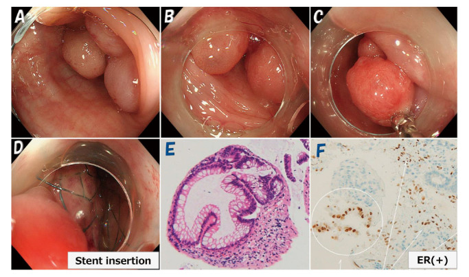
Polypoid endometriosis is a rare form of endometriosis characterized by polypoid masses that histologically often resemble endometrial polyps. We report a case of rapidly progressing polypoid endometriosis that was preoperatively assumed to be advanced ovarian cancer. A 46-year-old woman, para 0, underwent laparoscopic myomectomy and left adnexectomy for uterine fibroids and a left ovarian endometrial cyst after administration of gonadotropin releasing hormone (GnRH) agonist for 4 months. Eleven months postoperatively, rapid right ovarian enlargement occurred. CT and MRI (both contrast-enhanced) showed masses in the right adnexa, cecum, sigmoid colon, and omentum, and PET-CT demonstrated increased uptake, suggesting ovarian cancer and peritoneal dissemination. The patient later developed intestinal obstruction, and colonoscopy revealed multiple polypoid lesions in the sigmoid colon. The omental tumor and right adnexa were biopsied during exploratory laparotomy, and diagnosed as polypoid endometriosis with no malignancy by permanent pathology. The right adnexal tumor shrunk markedly after 4 months of GnRH antagonist treatment. Second laparotomy was then performed for right adnexal tumor resection and ileocecectomy. Pathological examination revealed polypoid endometriosis extending from the ovary to the cecal mucosa. The patient has been asymptomatic for over 1 year postoperatively. The sigmoid colon tumor shrunk but is still present.Polypoid endometriosis predominantly affects the ovaries, colon, peritoneum, and omentum of patients in their 40s and 50s. It is a benign disease but is often difficult to distinguish from malignancy preoperatively because it rapidly forms numerous solid lesions. Although polypoid endometriosis is rare, with no specific imaging findings, including it in a differential diagnosis may facilitate preoperative identification.
Keywords: PET-CT; differentiated diagnosis; endometriosis-associated malignancy; magnetic resonance imaging (MRI); polypoid endometriosis.
Conflict of interest statement
The authors have no conflicts of interest to declare.
Figures


References
-
- McCluggage WG. Endometriosis-related pathology: a discussion of selected uncommon benign, premalignant and malignant lesions, Histipathology, 76: 76-92, 2020. - PubMed
-
- Mostoufizadeh M and Secully RE. Malignant tumor arising in endometriosis. Clin Obstet Gynecol, 23: 951-963, 1980. - PubMed
-
- Yamada Y, Miyamoto T, Horiuchi A, Ayumi Ohya, Tanri Shiozawa. Polypoid endometriosis of the ovary mimicking ovarian carcinoma dissemination: A case report and literature review. Obstet Gynaecol Res, 40: 1426-1430, 2014. - PubMed
-
- Parker RL, Dadmanesg F, Young RH, Clement PB. Polypoid endometriosis: A clinicopathologic analysis of 24 cases and a review of the literature. Am J Surg Pathol, 28: 285-297, 2004. - PubMed
-
- Tsai C, Huang SH, Huang CY. Polypoid endometriosis – A rare entity of endometriosis mimicking ovarian cancer. Taiwan J Obstet Gyneco, 58: 328-329, 2019. - PubMed