

Correlation between Retinal Vascularization and Disease Aggressiveness in Amyotrophic Lateral Sclerosis
- PMID: 36289652
- PMCID: PMC9598742
- DOI: 10.3390/biomedicines10102390
Correlation between Retinal Vascularization and Disease Aggressiveness in Amyotrophic Lateral Sclerosis
Abstract
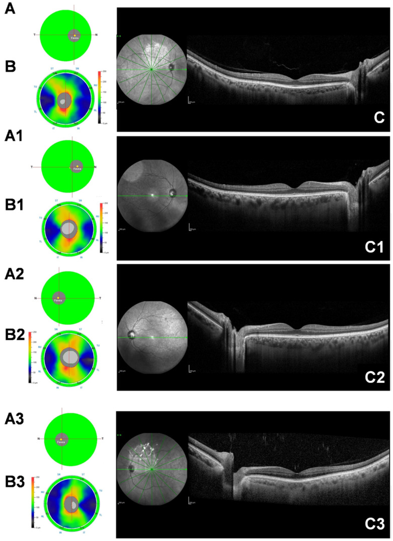
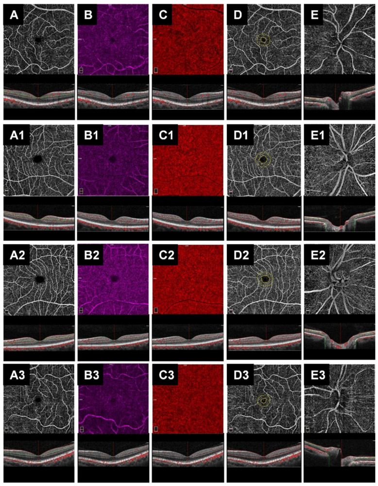
Abnormalities in retinal vascularization and neural density have been found in many neurodegenerative diseases; however, conflicting results are described in Amyotrophic Lateral Sclerosis (ALS). The aim of the present study was, therefore, to systematically analyze retinal layers and vascularization by means of spectral-domain (SD-OCT) and optical coherence tomography angiography (OCT-A) in ALS patients. We enrolled 48 ALS patients and 45 healthy controls. ALS patients were divided into three groups: slow progressors (n = 10), intermediate progressors (n = 24) and fast progressors (n = 14), according to the disease progression rate. For SD-OCT, we evaluated the Subfoveal choroidal thickness (SFCT), ganglion cell complex (GCC) and retinal nerve fiber layer (RNFL). Regarding the OCT-A, we assessed the vessel density (VD) in superficial and deep capillary plexuses, radial peripapillary capillary plexus, choriocapillary and the foveal avascular zone (FAZ) area. SD-OCT exam did not show any significant differences in GCC and RNFL thickness between patients and controls and among the three ALS groups. The SFCT was statistically greater in patients compared with controls (357.95 ± 55.15 µm vs. 301.3 ± 55.80 µm, p < 0.001); interestingly, the SFCT was thicker in patients with slow and intermediate disease progression than in those with fast disease progression (394.45 ± 53.73 µm vs. 393.09 ± 42.17 µm vs. 267.71 ± 56.24 µm, p < 0.001). OCT-A did not reveal any significant results. Amyotrophic Lateral Sclerosis Functional Rating Scale-Revised (ALSFRS-r) and disease duration did not correlate with any of the OCT parameters, except for SFCT with ALSFRS-r (r = 0.753, p = 0.024). This study demonstrated the possible association between choroidal thickness and disease activity in ALS. OCT could be a useful biomarker in the management of the disease.
Keywords: ALS; OCT; angiography; biomarker; choroid; disease progression; eye; inflammation; retinal nerve fiber layer; vascular.
Conflict of interest statement
The authors declare no conflict of interest. The funders had no role in the design of the study, in the collection, analyses or interpretation of data, in the writing of the manuscript or in the decision to publish the results.
Figures
References
Grants and funding
LinkOut - more resources
Full Text Sources
Miscellaneous
