Review
. 2022 Sep 27;10(10):2417.
doi: 10.3390/biomedicines10102417.

Advances in Musculoskeletal Imaging in Juvenile Idiopathic Arthritis

Affiliations
Review

Advances in Musculoskeletal Imaging in Juvenile Idiopathic Arthritis

Iwona Sudoł-Szopińska et al. Biomedicines. .

Abstract

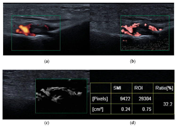
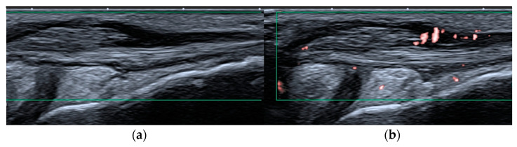
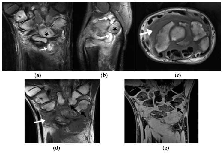
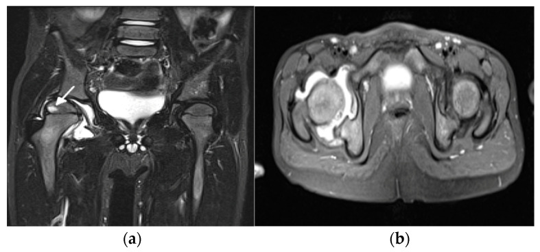
Over the past decade, imaging of inflammatory arthritis in juvenile arthropathies has significantly advanced due to technological improvements in the imaging modalities and elaboration of imaging recommendations and protocols through systematic international collaboration. This review presents the latest developments in ultrasound (US) and magnetic resonance imaging (MRI) of the peripheral and axial joints in juvenile idiopathic arthritis. In the field of US, the ultra-wideband and ultra-high-frequency transducers provide outstanding spatial resolution. The more sensitive Doppler options further improve the assessment and quantification of the vascularization of inflamed tissues, and shear wave elastography enables the diagnosis of tissue stiffness. Concerning MRI, substantial progress has been achieved due to technological improvements in combination with the development of semiquantitative scoring systems for the assessment of inflammation and the introduction of new definitions addressing the pediatric population. New solutions, such as superb microflow imaging, shear wave elastography, volume-interpolated breath-hold examination, and MRI-based synthetic computed tomography open new diagnostic possibilities and, at the same time, pose new challenges in terms of clinical applications and the interpretation of findings.

Keywords: elastography; juvenile idiopathic arthritis; magnetic resonance imaging; ultrasonography.


Conflict of interest statement

The authors declare no conflict of interest.

Figures

Figure 1
Figure 1
Third metatarso-phalangeal (MTP3, green square) joint synovitis shown on: power Doppler ultrasound (PDUS) (a), color superb microvascular imaging (SMI) (b), monochromatic SMI (c), and vascularity index (d); improved detection of hyperemia (vessels) achieved with SMI compared with PDUS.
Figure 2
Figure 2
Peroneal tenosynovitis in a 6-year-old girl. Long-axis power Doppler ultrasound image (green rectangles) of the peroneal tendons (a) shows no hyperemia, which is evident on the more sensitive superb microvascular imaging ultrasound image (b), consistent with active tenosynovitis.
Figure 3
Figure 3
A 14-year-old boy with JIA and wrist arthritis. Coronal T2 Turbo inversion recovery magnitude (TIRM) (a), sagittal proton density fat saturated (b) and axial T1-weighted time spin echo (TSE) (c) MR images show synovitis (white arrows) and bone marrow edema (stars); coronal T1 TSE (d) and T1 volume-interpolated breath-hold examination (VIBE) (e) show numerous cyst-like changes and erosions (black arrows).
Figure 4
Figure 4
A 5-year-old girl with juvenile idiopathic arthritis with hip involvement. Coronal short tau inversion recovery (STIR) (a) and axial proton density (PD)-weighted fat saturated (b) MR images show right hip effusion and bone marrow edema in the right femoral epiphysis (arrow).
Figure 5
Figure 5
A 1.5 Tesla temporomandibular joint MRI with T1- (a), fat-saturated T2- (b), and postcontrast fat-saturated T1-weighted images (c) in a 7-year-old girl with juvenile idiopathic arthritis showing active and chronic inflammatory lesions: synovitis (white arrow), bone marrow edema in the mandibular condyle (star) in the upper part of the ramus, and erosions (black arrow).
Figure 6
Figure 6
An 11-year-old old girl with juvenile idiopathic arthritis and cervical spine arthritis. Sagittal T1- (a), fat-saturated T2- (b), and postcontrast fat-saturated T1-weighted show prominent C1/2 joint arthritis with atlantodental joint fluid and synovial enhancement (arrows). Follow-up MRI with postcontrast fat-saturated T1-weighted sagittal MR image (c) 2 years later shows ankylosis of the atlanto-occipital joint, still with bone marrow edema consistent with ongoing active inflammation/osteitis (star) (d).
Figure 6
Figure 6
An 11-year-old old girl with juvenile idiopathic arthritis and cervical spine arthritis. Sagittal T1- (a), fat-saturated T2- (b), and postcontrast fat-saturated T1-weighted show prominent C1/2 joint arthritis with atlantodental joint fluid and synovial enhancement (arrows). Follow-up MRI with postcontrast fat-saturated T1-weighted sagittal MR image (c) 2 years later shows ankylosis of the atlanto-occipital joint, still with bone marrow edema consistent with ongoing active inflammation/osteitis (star) (d).
Figure 7
Figure 7
Whole-body MRI (WBMRI) in a 12-year-old boy with enthesitis-related arthritis (ERA) subtype of juvenile idiopathic arthritis (JIA). Superposition of findings of ERA and chronic recurrent multifocal osteomyelitis (CRMO) is seen on this WBMRI examination with STIR images of multiple body parts in multiple directions (ag). Findings of CRMO are presented by ill-defined flamed-shaped areas of increased inversion recovery (IR) signal seen in the bilateral proximal humeral metaphyses and distal clavicles (a, arrows), distal femoral and proximal tibial metaphyses and at a lesser extent, epiphyses (d,e), distal tibial metaphyses and epiphyses, and tarsal and proximal metatarsals (g). Findings of JIA-related enthesopathies are represented by IR signal noted in the bilateral greater trochanters (b, arrows) at the site of insertion of gluteus medius and minimus, along the sacroiliac joints (c), iliac, long arrows and sacral aspects, arrowhead; (c), normal anterior–superior iliac spine apophyses short arrows), proximal and distal attachments of patellar tendon (d, arrows), and at the insertion of the Achilles tendon in the posterior calcaneus (g, arrow).
Figure 8
Figure 8
Follow-up MRI of the sacroiliac joints (only anterior superior part is shown) in a 14-year-old boy with juvenile spondyloarthritis (SpA), coronal short tau inversion recovery (STIR) (a), T1 TSE (b), and volume-interpolated breath-hold examination (VIBE) (c): joints without active lesions (a), with numerous erosions in the right joint that are difficult to depict on the T1, and are better visualized on the VIBE sequence (arrows).
Figure 9
Figure 9
MRI of the sacroiliac joints of a 11-year-old boy with an active erosion in the left sacrum seen in consecutive sequences: T1-weighted time spin echo (TSE) (a), T2 short tau inversion recovery (STIR) (b), postgadolinium fat-saturated T1-weighted MR image (c), T1 volume-interpolated breath-hold examination (VIBE) (d), and BoneMRI (e, arrow).

References

    1. Ostrowska M., Gietka P., Mańczak M., Michalski E., Sudoł-Szopińska I. MRI Findings in Hip in Juvenile Idiopathic Arthritis. J. Clin. Med. 2021;10:5252. doi: 10.3390/jcm10225252. - DOI - PMC - PubMed
    1. Lovell D.J. Juvenile idiopathic arthritis: Clinical features. In: Klippel J.H., Stone J.H., Crofford L.J., White P.F., editors. Primer on the Rheumatic Diseases. Springer; New York, NY, USA: 2008. pp. 142–148.
    1. El-Azeem M.I.A., Taha H.A., El-Sherif A.M. Role of MRI in evaluation of hip joint involvement in juvenile idiopathic arthritis. Egypt. Rheumatol. 2012;34:75–82. doi: 10.1016/j.ejr.2012.03.001. - DOI
    1. Argyropoulou M., Fanis S.L., Xenakis T., Efremidis S.C., Siamopoulou A. The role of MRI in the evaluation of hip joint disease in clinical subtypes of juvenile idiopathic arthritis. Br. J. Radiol. 2002;75:229–233. doi: 10.1259/bjr.75.891.750229. - DOI - PubMed
    1. Murray J.G., Ridley N.T., Mitchell N., Rooney M. Juvenile chronic arthritis of the hip: Value of contrast-enhanced MR imaging. Clin. Radiol. 1996;51:99–102. doi: 10.1016/S0009-9260(96)80264-9. - DOI - PubMed

LinkOut - more resources