

Development of Real-Time Kinematic Magnetic Resonance Imaging (kMRI) Techniques for Studying the Kinematics of the Spine and Joints in Dogs-Preliminary Study on Cadavers
- PMID: 36290176
- PMCID: PMC9597841
- DOI: 10.3390/ani12202790
Development of Real-Time Kinematic Magnetic Resonance Imaging (kMRI) Techniques for Studying the Kinematics of the Spine and Joints in Dogs-Preliminary Study on Cadavers
Abstract
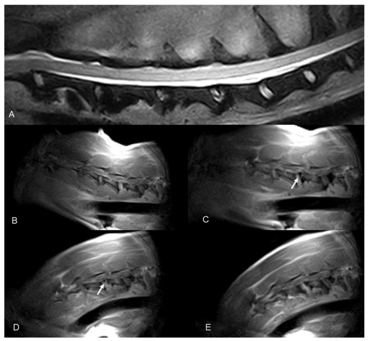
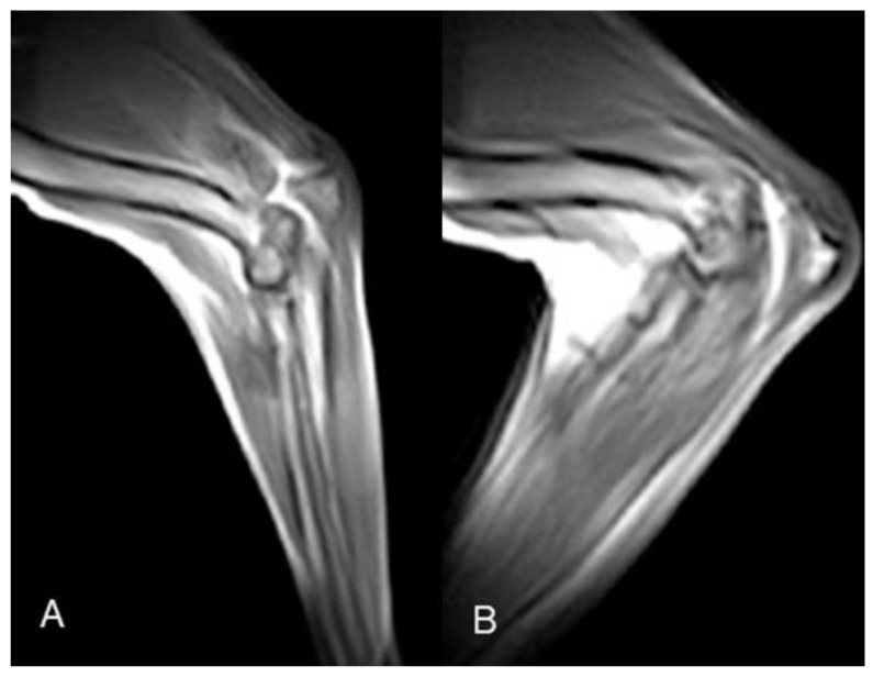
Kinematic MRI (kMRI) is a novel human imaging technique that couples the excellent soft tissue contrast and multiplanar capabilities of traditional MRI with kinematic potential. The study's goals are: (1) testing the feasibility of spinal cord and joints real-time kMRI; and (2) evaluating the quality of these kinematic studies as a new diagnostic option in veterinary medicine. Standard and real-time kinematic MRI were performed on cervical spine, elbow, and stifle joints of seven cadavers. Studies were repeated after a surgical insult aimed to create a certain degree of joint instability. A total of 56 MRI were performed-7 cervical spinal tracts, 3 elbow joints, and 4 stifle joints were examined. The technique was feasible in all the three regions examined. The images were considered of excellent quality for the stifle joint, good to fair for the cervical spine, whereas two of three elbow studies were considered to have unacceptable image quality. Additionally, real-time kMRI provided good to excellent information about stifle instability. Therefore we consider kMRI a promising technique in veterinary medicine. Further studies and an in vivo setting are needed to increase the quality of the kMRI images, and to fully evaluate clinical usefulness.
Keywords: canine; cervical spine; elbow; real-time kinematic MRI; stifle.
Conflict of interest statement
The authors declare no conflict of interest.
Figures


References
-
- Gavin P.R., Holmes S.P. Orthopedic. In: Gavin P.R., Bagley R.S., editors. Practical Small Animal MRI. Wiley-Blackwell; Ames, IA, USA: 2009. pp. 233–272.
LinkOut - more resources
Full Text Sources
