. 2022 Sep 20;11(10):1377.
doi: 10.3390/biology11101377.

Interleukin-10 Deficiency Impacts on TNF-Induced NFκB Regulated Responses In Vivo

Affiliations

Interleukin-10 Deficiency Impacts on TNF-Induced NFκB Regulated Responses In Vivo

Stamatia Papoutsopoulou et al. Biology (Basel). .

Abstract

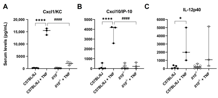
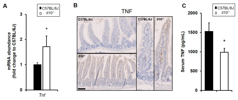
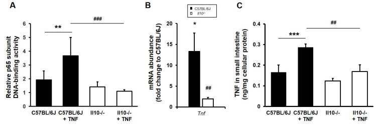
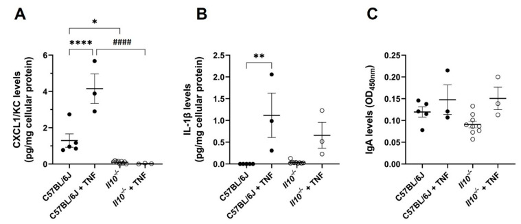
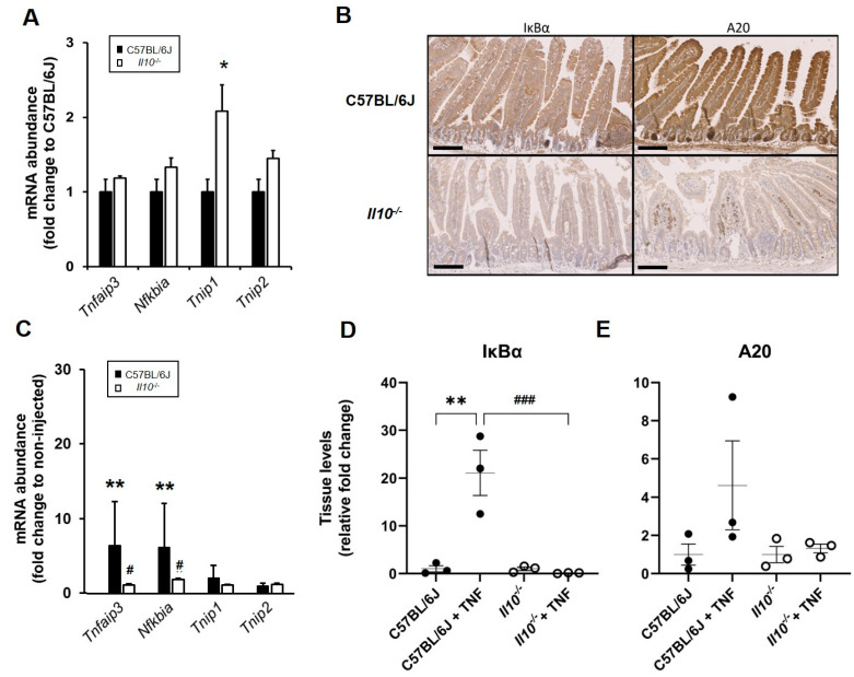
Interleukin-10 (IL-10) is an anti-inflammatory cytokine that has a major protective role against intestinal inflammation. We recently revealed that intestinal epithelial cells in vitro regulate NFκB-driven transcriptional responses to TNF via an autocrine mechanism dependent on IL-10 secretion. Here in this study, we investigated the impact of IL-10 deficiency on the NFκB pathway and its downstream targets in the small intestinal mucosa in vivo. We observed dysregulation of TNF, IκBα, and A20 gene and protein expression in the small intestine of steady-state or TNF-injected Il10-/- mice, compared to wild-type C57BL6/J counterparts. Upon TNF injection, tissue from the small intestine showed upregulation of NFκB p65[RelA] activity, which was totally diminished in Il10-/- mice and correlated with reduced levels of TNF, IκBα, and A20 expression. In serum, whilst IgA levels were noted to be markedly downregulated in IL-10-deficient- mice, normal levels of mucosal IgA were seen in intestine mucosa. Importantly, dysregulated cytokine/chemokine levels were observed in both serum and intestinal tissue lysates from naïve, as well as TNF-injected Il10-/- mice. These data further support the importance of the IL-10-canonical NFκB signaling pathway axis in regulating intestinal mucosa homeostasis and response to inflammatory triggers in vivo.

Keywords: NFκB; RelA; interleukin-10; intestine; mucosa; tumour necrosis factor.


Conflict of interest statement

All authors declare no competing commercial or financial interests in relation to the work described. The funders had no role in the design of the study; in the collection, analyses, or interpretation of data; in the writing of the manuscript; or in the decision to publish the results.

Figures

Figure 1
Figure 1
Attenuated serum levels of Cxcl1/KC and Cxcl10/IP-10 seen in IL-10-deficient mice following intra-peritoneal injection with TNF. Levels of (A) Cxcl1/KC, (B) Cxcl10/IP-10, and (C) IL-12p40 in serum of C57BL/6J (N = 5) and Il10−/− mice (N = 8) at resting levels, and those receiving i.p. injection of 0.33 mg TNF/kg body weight for 1.5 h (both genotypes N = 3). Serum was obtained from whole blood collected by cardiac puncture, with cytokines/chemokines (pg/mL) measured by ELISA. Significant differences compared to C57BL/6J control mice at rest, * p < 0.05, **** p < 0.0001; and significant differences from TNF-treated C57BL/6J mice, #### p < 0.0001 (ANOVA).
Figure 2
Figure 2
Dysregulated expression of TNF in resting small intestine of IL-10-deficient mice. C57BL/6J and Il10−/− mice under resting conditions were sacrificed, their sera collected, and tissue processed to assess for an abundance of mRNA and protein. (A) Enhanced Tnf mRNA levels were detected by qPCR in the small intestine of resting Il10−/− mice relative to C57BL/6J wild-type mice (both N = 3). (B) The C57BL/6J and Il10−/− small intestine was fixed in 4% w/v paraformaldehyde, with 4 μm sections processed and stained using antibodies against TNF. TNF staining appears only in lamina propria cells from Il10-deficient mice, but not in wild-type. Figures representative of N = 6 mice; three males and three females from each genotype; bar = 100 μm. (C) TNF levels in serum (pg/mL) were measured by ELISA, with lower levels being observed in IL-10-deficient mice under resting conditions (N = 8) compared to C57BL/6J (N = 5). Significant differences compared to C57BL/6J, * p < 0.05, Mann–Whitney U test.
Figure 3
Figure 3
Aberrant NFκB activation and inducible TNF expression in the small intestine of Il10-deficient mice. C57BL/6J and Il10−/− mice were either kept at resting conditions or following intraperitoneal injection with recombinant murine TNF (0.33 mg/kg body weight). Mice were sacrificed after 1.5 h and tissue processed to assess for NFκB signal pathway activation and for abundance of Tnf mRNA and TNF protein. (A) NFκB ELISA of small intestine tissue lysates revealed TNF-induced p65[RelA] transcriptional activation in wild-type mice, which was impaired in the tissue of IL-10-deficient mice. (B) TNF-induced Tnf transcript levels were observed to be significantly diminished in Il10−/− mice. (C) Similarly, tissue levels of TNF protein were not induced in intestinal mucosa of Il10−/− mice 1.5 h post TNF injection. All data are presented as mean ± SEM, N = 3 mice. Significant differences for TNF-injected versus resting levels in C57BL/6J mice, * p < 0.05, ** p < 0.01, and *** p < 0.001; and TNF-injected Il10−/− versus TNF-injected C57BL/6J, ## p < 0.01 and ### p < 0.001 (Kruskal–Wallis test or ANOVA).
Figure 4
Figure 4
Attenuated intestinal tissue levels of NFκB-regulated pro-inflammatory cytokines, but not mucosal IgA, seen in IL-10-deficient mice following intra-peritoneal injection with TNF. Levels of (A) CXCL1/KC, (B) IL-1β, and (C) IgA in small intestinal tissue lysates of C57BL/6J (N = 5) and Il10−/− mice (N = 9) at resting levels, and those who received an i.p. injection of 0.33 mg TNF per kg body weight for 1.5 h (both genotypes N = 3). Cytokine/chemokine levels were measured by ELISA. Mucosal IgA was measured using the Ig Isotyping mouse uncoated ELISA. There were significant differences compared to C57BL/6J control mice at rest, * p < 0.05, ** p < 0.01, and **** p < 0.0001; and significant differences from TNF-treated C57BL/6J mice, #### p < 0.0001 (ANOVA).
Figure 5
Figure 5
Dysregulated expression of key NFκB-targets in the small intestine of naïve and TNF-injected mice. C57BL/6J and Il10−/− mice were sacrificed at rest or following i.p. injection with recombinant murine TNF (0.33 mg/kg body weight). Tissue was dissected and processed for gene and protein expression analyses. (A) Expression of NFκB-target genes at rest in Il10−/− intestinal tissue (white bars) as assessed by qPCR. Data are presented as mean ± SEM, expressed as fold change to C57BL/6J (both N = 3 mice; significant difference compared to C57BL/6J, * p < 0.05 Kruskal–Wallis test). (B) Dysregulated A20 and IκBα protein expression in resting small intestine of Il10-deficient mice (as assessed by immunohistochemistry of 4% w/v paraformaldehyde-fixed, 4 μm microtomy tissue sections). Representative images from N = 6 mice; bar =100μm). (C) NFκB-target gene expression 1.5 h post-TNF injection of Il10−/− (white bars) compared to injected C57BL/6J mice (black bars); each N = 3 mice, with significant differences compared to non-injected controls ** p < 0.01, and to TNF-injected C57BL6/J # p < 0.01 (Kruskal–Wallis test). Intestinal tissue levels of (D) IκBα and (E) A20, expressed as relative fold change to respective genotype controls (N = 3 for all groups). There are significant differences compared to C57BL/6J * p < 0.05 and ** p < 0.01; and TNF-injected controls, ### p < 0.001 (ANOVA).

Similar articles

Cited by

References

    1. Wei H.X., Wang B., Li B. IL-10 and IL-22 in Mucosal Immunity: Driving Protection and Pathology. Front. Immunol. 2020;11:1315. doi: 10.3389/fimmu.2020.01315. - DOI - PMC - PubMed
    1. Kamanaka M., Kim S.T., Wan Y.Y., Sutterwala F.S., Lara-Tejero M., Galan J.E., Harhaj E., Flavell R.A. Expression of interleukin-10 in intestinal lymphocytes detected by an interleukin-10 reporter knockin tiger mouse. Immunity. 2006;25:941–952. doi: 10.1016/j.immuni.2006.09.013. - DOI - PubMed
    1. Bando J.K., Gilfillan S., Di Luccia B., Fachi J.L., Secca C., Cella M., Colonna M. ILC2s are the predominant source of intestinal ILC-derived IL-10. J. Exp. Med. 2020;217:e20191520. doi: 10.1084/jem.20191520. - DOI - PMC - PubMed
    1. Morhardt T.L., Hayashi A., Ochi T., Quiros M., Kitamoto S., Nagao-Kitamoto H., Kuffa P., Atarashi K., Honda K., Kao J.Y., et al. IL-10 produced by macrophages regulates epithelial integrity in the small intestine. Sci. Rep. 2019;9:1223. doi: 10.1038/s41598-018-38125-x. - DOI - PMC - PubMed
    1. Krause P., Morris V., Greenbaum J.A., Park Y., Bjoerheden U., Mikulski Z., Muffley T., Shui J.W., Kim G., Cheroutre H., et al. IL-10-producing intestinal macrophages prevent excessive antibacterial innate immunity by limiting IL-23 synthesis. Nat. Commun. 2015;6:7055. doi: 10.1038/ncomms8055. - DOI - PMC - PubMed

LinkOut - more resources