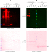
SNRPD2 Is a Novel Substrate for the Ubiquitin Ligase Activity of the Salmonella Type III Secretion Effector SlrP
- PMID: 36290420
- PMCID: PMC9598574
- DOI: 10.3390/biology11101517
SNRPD2 Is a Novel Substrate for the Ubiquitin Ligase Activity of the Salmonella Type III Secretion Effector SlrP
Abstract
SlrP is a protein with E3 ubiquitin ligase activity that is translocated by Salmonella enterica serovar Typhimurium into eukaryotic host cells through a type III secretion system. A yeast two-hybrid screen was performed to find new human partners for this protein. Among the interacting proteins identified by this screen was SNRPD2, a core component of the spliceosome. In vitro ubiquitination assays demonstrated that SNRPD2 is a substrate for the catalytic activity of SlrP, but not for other members of the NEL family of E3 ubiquitin ligases, SspH1 and SspH2. The lysine residues modified by this activity were identified by mass spectrometry. The identification of a new ubiquitination target for SlrP is a relevant contribution to the understanding of the role of this Salmonella effector.
Keywords: RNA splicing; SNRPD2; SlrP; type III secretion; ubiquitination.
Conflict of interest statement
The authors declare no conflict of interest. The funders had no role in the design of the study; in the collection, analyses, or interpretation of data; in the writing of the manuscript; or in the decision to publish the results.
Figures


Similar articles
-
Specificities and redundancies in the NEL family of bacterial E3 ubiquitin ligases of Salmonella enterica serovar Typhimurium.Front Immunol. 2024 Feb 1;15:1328707. doi: 10.3389/fimmu.2024.1328707. eCollection 2024. Front Immunol. 2024. PMID: 38361917 Free PMC article.
-
Salmonella type III secretion effector SlrP is an E3 ubiquitin ligase for mammalian thioredoxin.J Biol Chem. 2009 Oct 2;284(40):27587-95. doi: 10.1074/jbc.M109.010363. Epub 2009 Aug 18. J Biol Chem. 2009. PMID: 19690162 Free PMC article.
-
The Salmonella type III secretion effector, salmonella leucine-rich repeat protein (SlrP), targets the human chaperone ERdj3.J Biol Chem. 2010 May 21;285(21):16360-8. doi: 10.1074/jbc.M110.100669. Epub 2010 Mar 24. J Biol Chem. 2010. PMID: 20335166 Free PMC article.
-
The NEL Family of Bacterial E3 Ubiquitin Ligases.Int J Mol Sci. 2022 Jul 13;23(14):7725. doi: 10.3390/ijms23147725. Int J Mol Sci. 2022. PMID: 35887072 Free PMC article. Review.
-
Ubiquitination as an efficient molecular strategy employed in salmonella infection.Front Immunol. 2014 Nov 25;5:558. doi: 10.3389/fimmu.2014.00558. eCollection 2014. Front Immunol. 2014. PMID: 25505465 Free PMC article. Review.
Cited by
-
Specificities and redundancies in the NEL family of bacterial E3 ubiquitin ligases of Salmonella enterica serovar Typhimurium.Front Immunol. 2024 Feb 1;15:1328707. doi: 10.3389/fimmu.2024.1328707. eCollection 2024. Front Immunol. 2024. PMID: 38361917 Free PMC article.
-
Speaking the host language: how Salmonella effector proteins manipulate the host.Microbiology (Reading). 2023 Jun;169(6):001342. doi: 10.1099/mic.0.001342. Microbiology (Reading). 2023. PMID: 37279149 Free PMC article. Review.
-
Recent progress in molecular mechanisms of Salmonella effectors involved in gut epithelium invasion.Mol Biol Rep. 2025 Jun 16;52(1):601. doi: 10.1007/s11033-025-10715-9. Mol Biol Rep. 2025. PMID: 40522423 Review.
-
Trichinella spiralis inhibits myoblast differentiation by targeting SQSTM1/p62 with a secreted E3 ubiquitin ligase.iScience. 2024 Feb 2;27(3):109102. doi: 10.1016/j.isci.2024.109102. eCollection 2024 Mar 15. iScience. 2024. PMID: 38380253 Free PMC article.
-
Salmonella Type III Secretion System Effectors.Int J Mol Sci. 2025 Mar 14;26(6):2611. doi: 10.3390/ijms26062611. Int J Mol Sci. 2025. PMID: 40141253 Free PMC article. Review.
References
Grants and funding
- PID2019-106132RB-I00/AEI/10.13039/501100011033/Ministerio de Ciencia e Innovación - Agencia Estatal de Investigación
- P20_00576/Fondo Europeo de Desarrollo Regional (FEDER) y Consejería de Transformación Económica, Industria, Conocimiento y Universidades de la Junta de Andalucía
- US-1380805/Universidad de Sevilla, Fondo Europeo de Desarrollo Regional (FEDER) y Consejería de Transformación Económica, Industria, Conocimiento y Universidades de la Junta de Andalucía
LinkOut - more resources
Full Text Sources
Molecular Biology Databases

