. 2022 Oct 6;12(10):831.
doi: 10.3390/bios12100831.

An Exosomal miRNA Biomarker for the Detection of Pancreatic Ductal Adenocarcinoma

Affiliations

An Exosomal miRNA Biomarker for the Detection of Pancreatic Ductal Adenocarcinoma

Amy Makler et al. Biosensors (Basel). .

Abstract

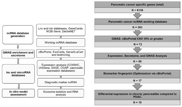
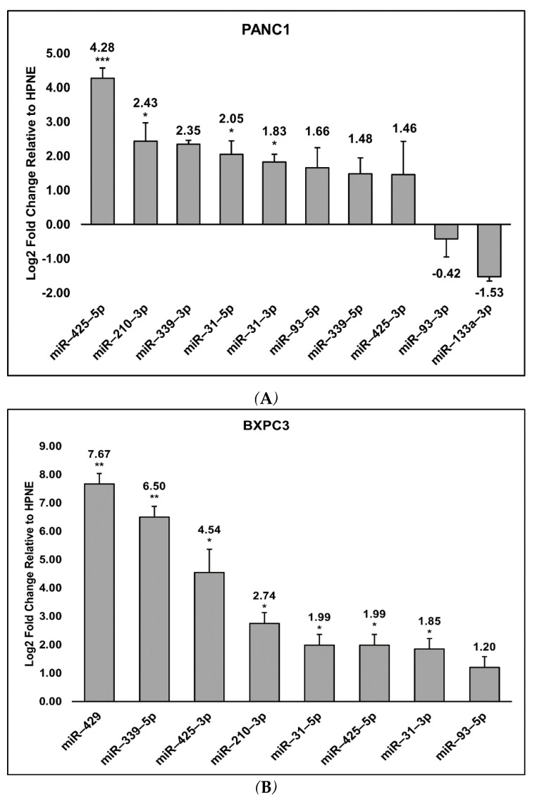
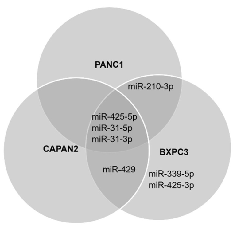
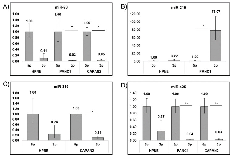
Pancreatic ductal adenocarcinoma (PDAC) remains a difficult tumor to diagnose and treat. To date, PDAC lacks routine screening with no markers available for early detection. Exosomes are 40-150 nm-sized extracellular vesicles that contain DNA, RNA, and proteins. These exosomes are released by all cell types into circulation and thus can be harvested from patient body fluids, thereby facilitating a non-invasive method for PDAC detection. A bioinformatics analysis was conducted utilizing publicly available miRNA pancreatic cancer expression and genome databases. Through this analysis, we identified 18 miRNA with strong potential for PDAC detection. From this analysis, 10 (MIR31, MIR93, MIR133A1, MIR210, MIR330, MIR339, MIR425, MIR429, MIR1208, and MIR3620) were chosen due to high copy number variation as well as their potential to differentiate patients with chronic pancreatitis, neoplasms, and PDAC. These 10 were examined for their mature miRNA expression patterns, giving rise to 18 mature miRs for further analysis. Exosomal RNA from cell culture media was analyzed via RTqPCR and seven mature miRs exhibited statistical significance (miR-31-5p, miR-31-3p, miR-210-3p, miR-339-5p, miR-425-5p, miR-425-3p, and miR-429). These identified biomarkers can potentially be used for early detection of PDAC.

Keywords: biomarker; cancer; diagnostics; exosomes; miRNA; pancreatic cancer.


Conflict of interest statement

The authors declare no conflict of interest.

Figures

Figure 1
Figure 1
Workflow for the identification and analysis of exosomal microRNA for PDAC diagnosis. Whole gene lists associated with PDAC were downloaded from the lnc and miRNA databases as well as GeneCards, NCBI Gene, and DisGenNET in order to identify as many PDAC-associated ncRNAs as possible (N = 6136). Protein-coding genes were then removed in order to isolate only ncRNAs (N = 383). This provided the basis for our ncRNA database. cBioPortal was used to identify genetic alterations of ncRNAs across patient data in order to determine the most attractive targets for diagnostic potential (N = 72). The expression databases (COSMIC, UniGene, SAGE, CGAP, and PED) and secretome tools (ExoCarta and GeneALaCart) were then accessed to determine if any of the remaining MIRs were secreted and exhibited changes in expression in PDAC to better determine the most reliable exosomal targets for PDAC detection (N = 50). Additional optimization using cBioPortal with putative secreted exosomal MIR markers (CNV 15% or greater) was used (N = 18). Comparison of MIR expression in chronic pancreatitis compared to PDAC was then used as the final metric for candidate MIRs for diagnostic potential (N = 10). The MIRs identified in this manner were considered diagnostic markers for further analysis in cell culture models.
Figure 2
Figure 2
Relative expression levels of MIRs detected in PANC1, BXPC3, and CAPAN2 compared to HPNE. The log2 fold changes of the relative expression levels of miRs in the three different PDAC cell lines, PANC1 (A), BXPC3 (B), and CAPAN2 (C) compared to HPNE was calculated. The log2 fold change was calculated via ΔΔCq values. Significant (p ≤ 0.05, *), very significant (p ≤ 0.01, **), and extremely significant (p ≤ 0.001, ***) are also noted and determined using the student’s t-test.
Figure 2
Figure 2
Relative expression levels of MIRs detected in PANC1, BXPC3, and CAPAN2 compared to HPNE. The log2 fold changes of the relative expression levels of miRs in the three different PDAC cell lines, PANC1 (A), BXPC3 (B), and CAPAN2 (C) compared to HPNE was calculated. The log2 fold change was calculated via ΔΔCq values. Significant (p ≤ 0.05, *), very significant (p ≤ 0.01, **), and extremely significant (p ≤ 0.001, ***) are also noted and determined using the student’s t-test.
Figure 3
Figure 3
miRNAs differentially expressed in at least one PDAC cell line. The figure exhibits the statistically significant miRs specific to each cell line and which of those were shared. Significant expression of miR-31-5p, miR-31-3p, and miR-425-5p was observed in all three PDAC cell lines. Meanwhile, CAPAN2 and BXPC3 both significantly expressed miR-429. miR-210-3p exhibited significance in the PANC1 and BXPC3 cell culture models while miR-339-5p and miR-425-3p were the only miRs to be significantly expressed in a single cell line, BXPC3.
Figure 4
Figure 4
Preference for 5p and 3p arm across PDAC cell lines and HPNE control. Each cell line was examined for a preference in 5p or 3p arm of the mature miRNA Expression levels were calculated relative to the 5p arm of each miR and displayed as fold change values. (A) miR-93-5p was preferred in PANC1 and CAPAN2, while (B) miR-210-3p was preferred in PANC1. (C) miR-339-5p was preferred in CAPAN2 and (D) miR-425-5p were preferred in both PANC1 and CAPAN2. Significant (p ≤ 0.05, *) and very significant (p ≤ 0.01, **) are also noted and were determined using the student’s t-test.

References

    1. Balachandran V.P., Beatty G.L., Dougan S.K. Broadening the Impact of Immunotherapy to Pancreatic Cancer: Challenges and Opportunities. Gastroenterology. 2019;156:2056–2072. doi: 10.1053/j.gastro.2018.12.038. - DOI - PMC - PubMed
    1. Rahib L., Smith B.D., Aizenberg R., Rosenzweig A.B., Fleshman J.M., Matrisian L.M. Projecting Cancer Incidence and Deaths to 2030: The Unexpected Burden of Thyroid, Liver, and Pancreas Cancers in the United States. Cancer Res. 2014;74:2913–2921. doi: 10.1158/0008-5472.CAN-14-0155. - DOI - PubMed
    1. Hall B.R., Cannon A., Atri P., Wichman C.S., Smith L.M., Ganti A.K., Are C., Sasson A.R., Kumar S., Batra S.K. Advanced Pancreatic Cancer: A Meta-Analysis of Clinical Trials over Thirty Years. Oncotarget. 2018;9:19396–19405. doi: 10.18632/oncotarget.25036. - DOI - PMC - PubMed
    1. Guillén-Ponce C., Blázquez J., González I., De-Madaria E., Montáns J., Carrato A. Diagnosis and Staging of Pancreatic Ductal Adenocarcinoma. Clin. Transl. Oncol. 2017;19:1205–1216. doi: 10.1007/s12094-017-1681-7. - DOI - PubMed
    1. Gbolahan O.B., Tong Y., Sehdev A., O’Neil B., Shahda S. Overall Survival of Patients with Recurrent Pancreatic Cancer Treated with Systemic Therapy: A Retrospective Study. BMC Cancer. 2019;19:468. doi: 10.1186/s12885-019-5630-4. - DOI - PMC - PubMed

MeSH terms