. 2022 Oct 6;12(10):834.
doi: 10.3390/bios12100834.

Comparison between Chest-Worn Accelerometer and Gyroscope Performance for Heart Rate and Respiratory Rate Monitoring

Affiliations

Comparison between Chest-Worn Accelerometer and Gyroscope Performance for Heart Rate and Respiratory Rate Monitoring

Chiara Romano et al. Biosensors (Basel). .

Abstract

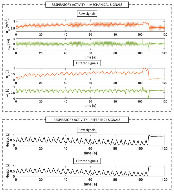
The demand for wearable devices to simultaneously monitor heart rate (HR) and respiratory rate (RR) values has grown due to the incidence increase in cardiovascular and respiratory diseases. The use of inertial measurement unit (IMU) sensors, embedding both accelerometers and gyroscopes, may ensure a non-intrusive and low-cost monitoring. While both accelerometers and gyroscopes have been assessed independently for both HR and RR monitoring, there lacks a comprehensive comparison between them when used simultaneously. In this study, we used both accelerometers and gyroscopes embedded in a single IMU sensor for the simultaneous monitoring of HR and RR. The following main findings emerged: (i) the accelerometer outperformed the gyroscope in terms of accuracy in both HR and RR estimation; (ii) the window length used to estimate HR and RR values influences the accuracy; and (iii) increasing the length over 25 s does not provide a relevant improvement, but accuracy improves when the subject is seated or lying down, and deteriorates in the standing posture. Our study provides a comprehensive comparison between two promising systems, highlighting their potentiality for real-time cardiorespiratory monitoring. Furthermore, we give new insights into the influence of window length and posture on the systems' performance, which can be useful to spread this approach in clinical settings.

Keywords: heart rate; magneto-inertial measurement units; mechanical vibrations; respiratory rate; wearable systems.


Conflict of interest statement

The authors declare no conflict of interest.

Figures

Figure 1
Figure 1
(a) Schematic illustration of the positioning of the IMU sensor (embedding the ACC and the GYR) on the subject’s chest from the frontal view. (b) Lateral view of the IMU sensor on the subject’s chest. Note that the sensor is integral with the body of the subject. This ensures that acceleration and angular velocity measurements correspond with good approximation to those experienced by the chest in response to cardiac and respiratory activity. (c) Example of an acceleration (in orange) and angular velocity (in green) signal filtered so that only the cardiac component is reported. (d) Example of an acceleration (in orange) and angular velocity (in green) signal filtered so that only the respiratory component is reported.
Figure 2
Figure 2
(a) Positioning of the IMU sensor at the fifth-left intercostal space in the midclavicular line of the mitral valve and of the wearable chest multiparametric device used as a reference system for registering both the ECG waveform and the respiratory waveform. (b) Picture of one subject during the protocol performed in the 3 at-rest postures, i.e., sitting, standing, and lying down.
Figure 3
Figure 3
Signal processing for cardiac activity extraction for mechanical signals (i.e., accelerations, a and angular velocities, ω) and reference signal (ECG). After acquiring the raw signals of the ACC and the GYR, only one axis was selected to reduce the dimensionality of the problem (i.e., the z axis for the ACC and the y axis for the GYR). The selected signals were reconstructed using the inverse wavelet transform by selecting frequencies between 10 Hz and 40 Hz and normalized. Finally, the normalized signal envelope was extracted to emphasize the heartbeats and filtered between 0.7 and 3 Hz. Concurrently, the raw ECG reference signal was filtered between 0.7 and 3 Hz.
Figure 4
Figure 4
Signal processing for respiratory activity extraction for mechanical signals (i.e., accelerations, a and angular velocities, ω) and reference respiratory waveform.
Figure 5
Figure 5
(a,d) After signal preprocessing, HR and RR values were extracted from both mechanical and reference signals using a frequency domain analysis in sliding windows. (b,e) In each window, the PSD was computed using Welch’s method and the dominant frequency was selected. This analysis was computed using six different window sizes. (c,f) An example for one subject of the HR and RR values extracted in all the window sizes is also reported.
Figure 6
Figure 6
Bland–Altman plot for HR estimation considering all postures from both a (in orange) and ω (in green) signals against reference. The dotted line in the figure represents the MOD, while the solid lines represent the LOA values. In each graph, the number of points is equal to the number of subjects multiplied by 120 (i.e., the duration of the test) minus the length of the window. For example, for the 5 s window, there will be 1256 points for a and 1256 points for ω.
Figure 7
Figure 7
Bar graph of the mean absolute error (MAE) values calculated for both HR analysis (in the upper graph) and RR analysis (in the bottom graph) and for the ACC (in orange) and the GYR (in green). Moreover, these values are reported for each subject tacking on the different postures (i.e., sitting, lying down, and standing) and for all the window lengths. For each analyzed combination, we included the mean MAE value (in black) considering all subjects and the relative standard deviations (reported as an error bar in red).
Figure 8
Figure 8
Bland–Altman plot for RR estimation considering all postures from both a (in orange) and ω (in green) signals against reference. The dashed line in the figure represents the MOD while the solid lines represent the LOA values. In each graph, the number of points is equal to the number of subjects multiplied by 120 (i.e., the duration of the test) minus the length of the window.

References

    1. Brüser C., Antink C.H., Wartzek T., Walter M., Leonhardt S. Ambient and Unobtrusive Cardiorespiratory Monitoring Techniques. IEEE Rev. Biomed. Eng. 2015;8:30–43. doi: 10.1109/RBME.2015.2414661. - DOI - PubMed
    1. Markova V., Ganchev T., Kalinkov K., Markov M. Detection of Acute Stress Caused by Cognitive Tasks Based on Physiological Signals. Bull. Electr. Eng. Inform. 2021;10:2539–2547. doi: 10.11591/eei.v10i5.3130. - DOI
    1. Tipton M.J., Harper A., Paton J.F.R., Costello J.T. The Human Ventilatory Response to Stress: Rate or Depth? J. Physiol. 2017;595:5729–5752. doi: 10.1113/JP274596. - DOI - PMC - PubMed
    1. Järvelin-Pasanen S., Sinikallio S., Tarvainen M.P. Heart Rate Variability and Occupational Stress—Systematic Review. Ind. Health. 2018;56:500–511. doi: 10.2486/indhealth.2017-0190. - DOI - PMC - PubMed
    1. Nicolò A., Massaroni C., Schena E., Sacchetti M. The Importance of Respiratory Rate Monitoring: From Healthcare to Sport and Exercise. Sensors. 2020;20:6396. doi: 10.3390/s20216396. - DOI - PMC - PubMed

LinkOut - more resources