. 2022 Oct 10;12(10):853.
doi: 10.3390/bios12100853.

All-Polymeric Electrode Based on PEDOT:PSS for In Vivo Neural Recording

Affiliations

All-Polymeric Electrode Based on PEDOT:PSS for In Vivo Neural Recording

Gilberto Filho et al. Biosensors (Basel). .

Abstract

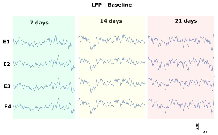
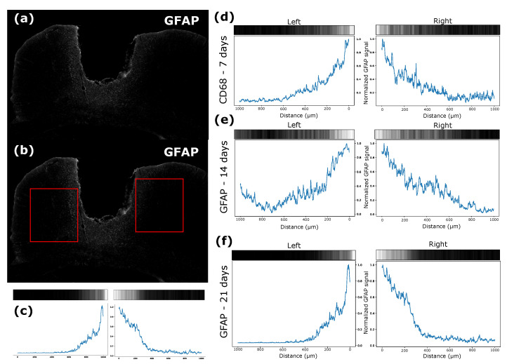
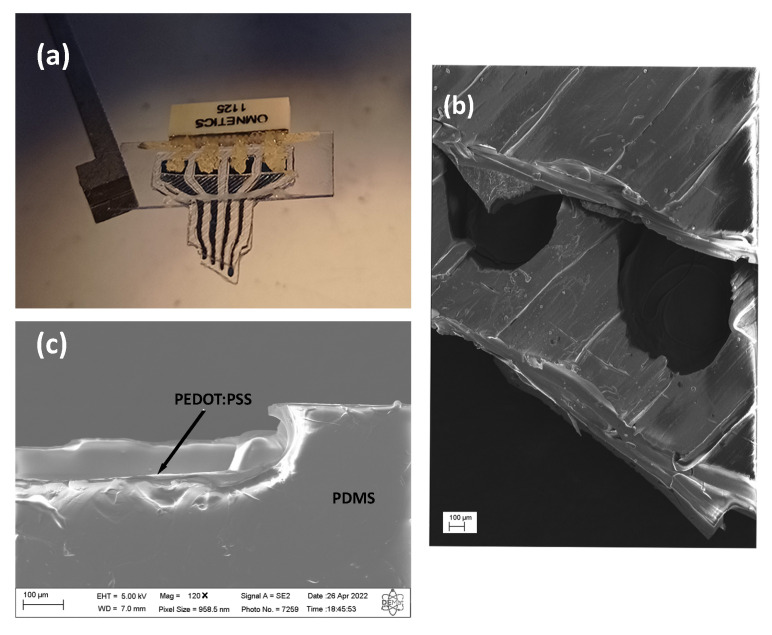
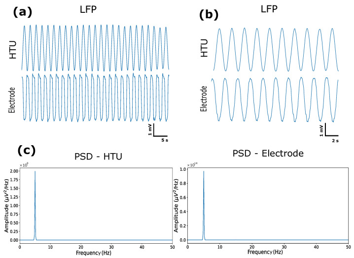
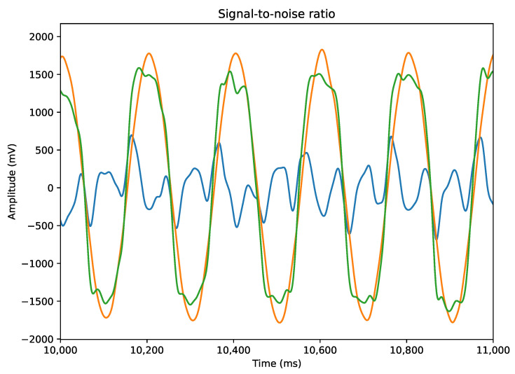
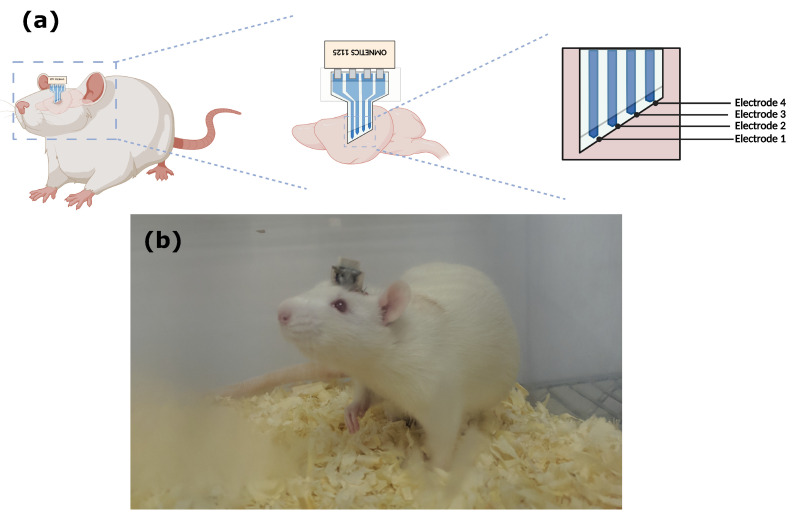
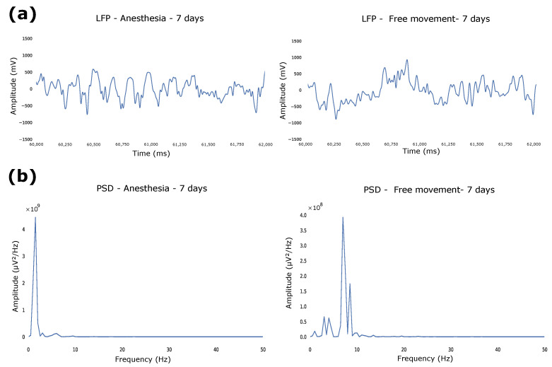
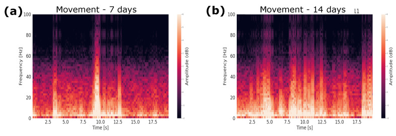
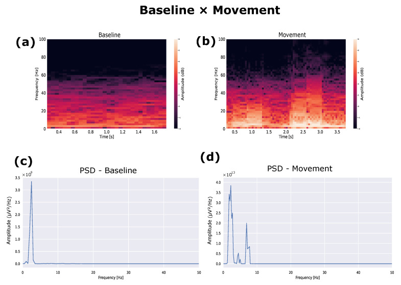
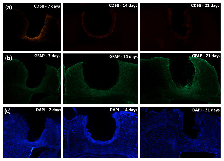
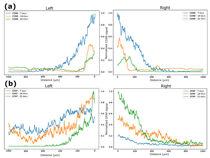
One of the significant challenges today in the brain-machine interfaces that use invasive methods is the stability of the chronic record. In recent years, polymer-based electrodes have gained notoriety for achieving mechanical strength values close to that of brain tissue, promoting a lower immune response to the implant. In this work, we fabricated fully polymeric electrodes based on PEDOT:PSS for neural recording in Wistar rats. We characterized the electrical properties and both in vitro and in vivo functionality of the electrodes. Additionally, we employed histological processing and microscopical visualization to evaluate the tecidual immune response at 7, 14, and 21 days post-implant. Electrodes with 400-micrometer channels showed a 12 dB signal-to-noise ratio. Local field potentials were characterized under two conditions: anesthetized and free-moving. There was a proliferation of microglia at the tissue-electrode interface in the early days, though there was a decrease after 14 days. Astrocytes also migrated to the interface, but there was not continuous recruitment of these cells in the tissue; there was inflammatory stability by day 21. The signal was not affected by this inflammatory action, demonstrating that fully polymeric electrodes can be an alternative means to prolong the valuable time of neural recordings.

Keywords: BMI; PEDOT:PSS; immune response; neural recording.


Conflict of interest statement

The authors declare no conflict of interest.

Figures

Figure 8
Figure 8
Analysis of functioning and intensity through the LFP of days 7, 14, and 21 of all channels.
Figure 11
Figure 11
(a) Image after treatment with averaging of the intensity vectors. (b) Quadrant used to measure GFAP intensity. (c) Signal strength near the electrode–tissue interface. (d) GFAP signal strength near the electrode–tissue interface after 7 days. (e) GFAP signal strength near the electrode–tissue interface after 14 days. (f) GFAP signal strength near the electrode–tissue interface after 21 days.
Figure 1
Figure 1
(a) Intracortical electrode. (b) Image of the intracortical channel obtained with SEM. (c) Interface deposition with PEDOT:PSS:DMSO and PDMS, layer deposition of 10.5 ± 1.2 μm.
Figure 2
Figure 2
(a) Artificial signal LFP with 5 s for the HTU and electrode. (b) Artificial signal LFP with 2 s for the HTU and electrode. (c) Artificial signal PSD with the HTU and electrode.
Figure 3
Figure 3
Difference between the signals detected by the HTU and by the electrode, and the influence of noise.
Figure 4
Figure 4
(a) Demonstration of the electrode in the rat brain (created with BioRender.com). (b) Wistar rat two days after surgery.
Figure 5
Figure 5
(a) LFP between anesthetized and free-movement conditions. (b) PSD between anesthetized and free-movement conditions.
Figure 6
Figure 6
Spectrogram of the moving rat: (a) 7 days and (b) 14 days.
Figure 7
Figure 7
(a) Spectrogram of the baseline rat. (b) Spectrogram of the moving rat. (c) PSD of the baseline rat. (d) PSD of the baseline rat during movement.
Figure 9
Figure 9
Tissue immune response to implant. (a) CD68-labeled cells at days 7, 14, and 21. (b) GFAP-labeled cells at days 7, 14, and 21. (c) DAPI-labeled cells at days 7, 14, and 21.
Figure 10
Figure 10
(a) Image after treatment with averaging of the intensity vectors. (b) Quadrant used to measure CD68 intensity. (c) Signal strength near the electrode–tissue interface. (d) CD68 signal strength near the electrode–tissue interface after 7 days. (e) CD68 signal strength near the electrode–tissue interface after 14 days. (f) CD68 signal strength near the electrode–tissue interface after 21 days.
Figure 12
Figure 12
(a) Normalized CD68 intensity over 7, 14, and 21 days. (b) Normalized GFAP intensity over 7, 14, and 21 days.

References

    1. Sohal H.S., Jackson A., Jackson R., Clowry G.J., Vassilevski K., O’Neill A., Baker S.N. The sinusoidal probe: A new approach to improve electrode longevity. Front. Neuroeng. 2014;7:10. doi: 10.3389/fneng.2014.00010. - DOI - PMC - PubMed
    1. Buzsáki G., Anastassiou C.A., Koch C. The origin of extracellular fields and currents—EEG, ECoG, LFP and spikes. Nat. Rev. Neurosci. 2012;13:407–420. doi: 10.1038/nrn3241. - DOI - PMC - PubMed
    1. Polikov V.S., Tresco P.A., Reichert W.M. Response of brain tissue to chronically implanted neural electrodes. J. Neurosci. Methods. 2005;148:1–18. doi: 10.1016/j.jneumeth.2005.08.015. - DOI - PubMed
    1. Gulino M., Kim D., Pané S., Santos S.D., Pêgo A.P. Tissue response to neural implants: The use of model systems toward new design solutions of implantable microelectrodes. Front. Neurosci. 2019;13:689. doi: 10.3389/fnins.2019.00689. - DOI - PMC - PubMed
    1. Kubota Y. Untangling GABAergic wiring in the cortical microcircuit. Curr. Opin. Neurobiol. 2014;26:7–14. doi: 10.1016/j.conb.2013.10.003. - DOI - PubMed

LinkOut - more resources