Review
. 2022 Oct 13;12(10):870.
doi: 10.3390/bios12100870.

Application of Microfluidics in Drug Development from Traditional Medicine

Affiliations
Review

Application of Microfluidics in Drug Development from Traditional Medicine

Xue Li et al. Biosensors (Basel). .

Abstract

While there are many clinical drugs for prophylaxis and treatment, the search for those with low or no risk of side effects for the control of infectious and non-infectious diseases is a dilemma that cannot be solved by today's traditional drug development strategies. The need for new drug development strategies is becoming increasingly important, and the development of new drugs from traditional medicines is the most promising strategy. Many valuable clinical drugs have been developed based on traditional medicine, including drugs with single active ingredients similar to modern drugs and those developed from improved formulations of traditional drugs. However, the problems of traditional isolation and purification and drug screening methods should be addressed for successful drug development from traditional medicine. Advances in microfluidics have not only contributed significantly to classical drug development but have also solved many of the thorny problems of new strategies for developing new drugs from traditional drugs. In this review, we provide an overview of advanced microfluidics and its applications in drug development (drug compound synthesis, drug screening, drug delivery, and drug carrier fabrication) with a focus on its applications in conventional medicine, including the separation and purification of target components in complex samples and screening of active ingredients of conventional drugs. We hope that our review gives better insight into the potential of traditional medicine and the critical role of microfluidics in the drug development process. In addition, the emergence of new ideas and applications will bring about further advances in the field of drug development.

Keywords: bioMEMS; drug development; microfluidics; traditional medicine.


Conflict of interest statement

The authors declare no conflict of interest.

Figures

Figure 1
Figure 1
Schematic diagram of drug synthesis by Microchannel reactor: (A) Schematic diagram of the biosynthesis of ibuprofen in a microchannel reactor from [26], Copyright 2021, American Chemical Society. (B) Schematic diagram of the biosynthesis of isoquercitrin in a microchannel reactor from [35], Copyright 2017, Nature Publishing Group.
Figure 2
Figure 2
Schematic representation of the synthesis of active molecules with the potential to inhibit thrombin by a Droplet microreactor: (A) Schematic diagram of the reagent A droplets. (B) Schematic diagram of chemical synthesis by injection of microdroplets. (C) Schematic representation of the synthesis of molecules with potential prothrombin-inhibiting activity by amines, aldehydes, and isocyanides. The image from [38], Copyright 2012, Royal Society of Chemistry.
Figure 3
Figure 3
Schematic diagram of the Microfluidic chip for obtaining single cells: (A) Schematic diagram of the design of the Microfluidic chip. (B) Actual image of the Microfluidic chip, measuring 6.0 cm × 5.0 cm. (C) Schematic diagram of the operation of single cell sorting and reagent infusion. (D) Schematic diagram of the typical dynamic processes of single cells captured by (L1–L5) or passed through (S1–S5) filter cells. The image from [39], Copyright 2016, Royal Society of Chemistry.
Figure 4
Figure 4
Schematic diagram of drug screening by Droplet Microfluidic Chip: (A) Inject droplets containing cells and fluorescent dye. (B) Droplet merging is controlled by electricity. (C) Mixing dyes with cells. (D) Optimize cell staining by extending channels. (E) Collect fluorescent signals. The image from [46], Copyright 2009, PNAS.
Figure 5
Figure 5
Schematic diagram of a Microfluidic chip driven by pressure: (A) Eight different dyes are injected into the Microfluidic chip. (B) Pressure is applied by venting from the outside through a sterile ventilation filter, with the dashed line indicating the enlarged area in (C). (C) Microscopic photograph of the Microfluidic chip, with the dashed square indicating the expanded region in (D). (D) Three colors indicate three different microstructures. The image from [42], Copyright 2008, Wiley.
Figure 6
Figure 6
Schematic diagram of single-cell encapsulation in droplets by Microfluidic chip: (A) Microfluidic chip, arrows are droplet generation junctions, and boxes indicate droplet docking arrays. (A) Microfluidic chip, arrows are droplet generation junctions, and boxes indicate droplet docking arrays. (B) Droplet generation. (C) Microdroplet docking. (D) Detection of Cy-5 conjugated ABCB-1 mRNA in live MCF-7S cells in droplet (enlarged in inset), Hoechst labeled cells nuclei. (E) Dox-resistant MCF-7R cell encapsulation in droplet. Inset: Calcein AM localization in vesicles. The image from [47], Copyright 2015, Royal Society of Chemistry.
Figure 7
Figure 7
Schematic diagram of the creation of a three-dimensional scaffold in a droplet (composed of a water nucleus and a hydrogel shell): (A) Principle of obtaining alginate networks. (B) PDMS Microfluidic chip. (C) Alginate cross-linking in the Microfluidic chip. (D) Fluorescein-labeled alginate as shown in the inset where the shell of alginate hydrogel was found under confocal microscopy. The image from [48], Copyright 2016, Royal Society of Chemistry.
Figure 8
Figure 8
Schematic of a human on a microfluidic chip: (A) An integrated system composed of micro-organs. The image from [51], Copyright 2011, Elsevier. (B) Integrated system of multiple culture dishes connected by perfusable vessels. The image from [52], Copyright 2015, SAGE. (C) Integrated system of different organ-on-a-chip couplings. The image from [53], Royal Society of Chemistry.
Figure 9
Figure 9
Schematic diagram of the extraction of olive bitter glycosides by Microfluidics: (A) Microfluidic device. (B) Chromatogram of extracted olive bitter glucoside into aqueous phase: microchannel device (Ba) and ethyl acetate extract (Bb). The image from [5], Copyright 2018, Herbal Medicines Journal.
Figure 10
Figure 10
Schematic diagram of the microfluidic chip separating free compounds: three-phase microfluidic chip (A) and two-phase microfluidic chip (B). The image from [11], Copyright 2020, Elsevier.
Figure 11
Figure 11
Schematic diagram of the isolation of Scutellaria baicalensis extract by IPSE chip to obtain aglycones and glycosides: (A) IPSE chip. (Aa) IPSE chip when filled with dye mixture (indigo and Sudan red); (Ab) IPSE chip when using dichloromethane-butyl acetate (2:8) to separate the dye mixture. (B) HPLC profiles: baicalin (peak 11), wogonoside (peak 12), baicalein (peak 13), wogonin (peak 14). The image from [10], Copyright 2021, MDPI.
Figure 12
Figure 12
Schematic diagram of biopharmaceutical quality assessment and screening on a Microfluidic chip. The image from [2], Copyright 2017, Scientific Reports.
Figure 13
Figure 13
Schematic diagram of the layout and instrument setup of the Microfluidic chip: (A) Schematic diagram of the Microfluidic chip. (B) Schematic diagram of the instrument setup. (C) Single cells were fixed with a scale bar of 50 mm. The inset shows the fluorescence image of a RAW cell (7 mm in diameter) after stimulation with 10 mg/mL of ionomycin. The image from [14], Copyright 2009, The Royal Society of Chemistry.
Figure 14
Figure 14
Schematic representation of cisplatin, dinatin, diosmetin activity, and toxicity screening by Microfluidic chip: (A) Schematic diagram of the chip. (B) The calculated survival rate of cisplatin, dinatin, and diosmetin to HEK293 cell, along with the results of each compatibility group. The image from [16], Copyright 2019, Elsevier.
Figure 15
Figure 15
(Aa) Schematic diagram of the microfluidic system. (Ab) Verification of the integrity of the endothelial cell monolayer between collagen and HUVEC channels by immunofluorescence staining for VE-cadherin (green in the image). (Ac) List of 12 compounds tested in this study. (Ba) Images of A549 cancer spheroids before and after 36 h of treatment with various drugs. (Bb) Relative dispersion rates of spheroids in each sample group. (Bc) Relative dispersion rates of spheroids under the effect of different concentrations of compound 7. (Bd) Representative images of HUVECs prearranged along the channel (Calcein-AM staining in green) after 36 h of treatment with various chemical drugs, with compound 2 causing significant damage to the endothelium. The image from [18], Copyright 2014, American Chemical Society.
Figure 16
Figure 16
(Aa) Schematic diagram of the microfluidic chip. (Ab) Physical diagram of the microfluidic chip. (Ac) Schematic diagram of the working principle of the analyzed system. (Ad) Photograph of the analytical system. (B) Histogram of platelet surface coverage for different concentrations of pro-catechin. The image from [22], Copyright 2021, Hindawi.
Figure 17
Figure 17
(Aa) Structural view of the Microfluidic compact disc (CD). (Ab) Top view of the third layer of the antioxidant microfluidic chip. (Ac) The whole experimental setup. (B) Image and schematic diagram of the whole process on CD. (Ba) Initial of the experiment, speed is 0. (Bb) The plant extract starts to flow into the capillary valve, speed increases from 0 to 300 rpm. (Bc) The plant extract chamber is emptied, speed 300 rpm. (Bd) The DPPH solution flows from its reaction chamber to the capillary valve, speed increases slowly from 300 to 800 rpm. (Be) The DPPH reaction chamber is emptied, speed 800 rpm. (Bf) All solutions were properly mixed in the reaction chamber at a speed of 1400 rpm. (C) Comparison of the conventional and LoD methods at different antioxidant activities. The image from [67], Copyright 2018, MDPI.
Figure 18
Figure 18
(Aa) Schematic diagram of the LOAD layout. (Ab) Chambers, channels, valves and siphons for testing allergenicity in one unit. (Ac) A zoom-in diagram showing the flange in C5. (Ad) Assembly of the integrated microfluidic layer. (Ba) Schematic diagram of AO as a degranulation reporter. (Bb) Red-shift phenomenon. (C) Triptolide suppressed the fMLP-mediated AO release in KU-812 cell. The image from [68], Copyright 2016, MDPI.

Similar articles

Cited by

References

    1. Thomford N., Senthebane D., Rowe A., Munro D., Seele P., Maroyi A., Dzobo K. Natural Products for Drug Discovery in the 21st Century: Innovations for Novel Drug Discovery. Int. J. Mol. Sci. 2018;19:1578. doi: 10.3390/ijms19061578. - DOI - PMC - PubMed
    1. Li Z., Ai N., Yu L., Qian Z., Cheng Y. A multiple biomarker assay for quality assessment of botanical drugs using a versatile microfluidic chip. Sci. Rep. 2017;7:12243. doi: 10.1038/s41598-017-12453-w. - DOI - PMC - PubMed
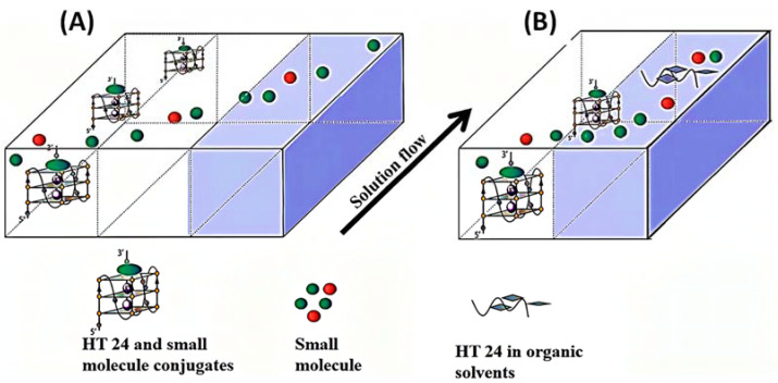
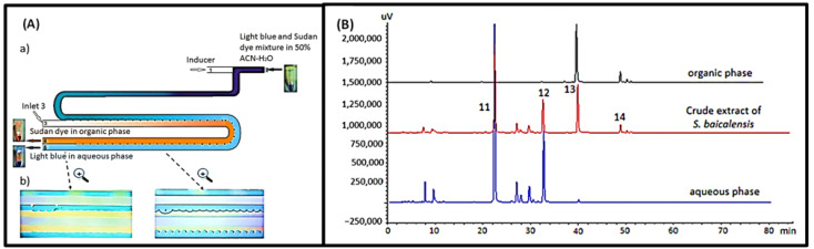
    1. Qin W., He Y., Xiao J., Liang S., Wang S., Li P.C.H., Sun Y. A successive laminar flow extraction for plant medicine preparation by microfluidic chip. Microfluid. Nanofluidics. 2019;23:61. doi: 10.1007/s10404-019-2228-8. - DOI
    1. Fan J., Bao Y., Meng X., Wang S., Li T., Chang X., Yang G., Bo T. Mechanism of modulation through PI3K-AKT pathway about Nepeta cataria L.’s extract in non-small cell lung cancer. Oncotarget. 2017;8:31395–31405. doi: 10.18632/oncotarget.15608. - DOI - PMC - PubMed
    1. Wei R., Mi D., Yang L., Cui Y. Research Progress of Traditional Chinese Medicine Treatment on DiabeticNephropathy. TCM. 2022;35:82–86.

LinkOut - more resources