. 2022 Oct 20;11(20):3297.
doi: 10.3390/cells11203297.

Modulation of Neutrophil Activity by Soluble Complement Cleavage Products-An In-Depth Analysis

Affiliations

Modulation of Neutrophil Activity by Soluble Complement Cleavage Products-An In-Depth Analysis

Lisa Wohlgemuth et al. Cells. .

Abstract

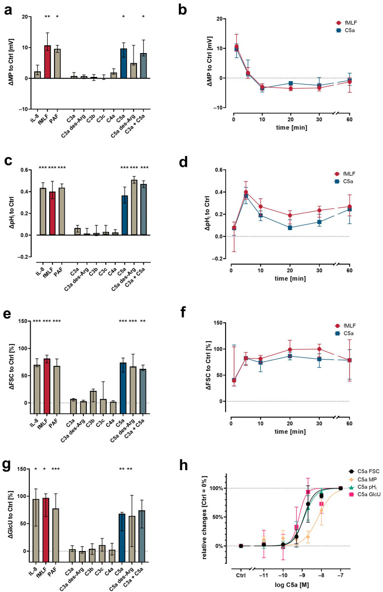
The cellular and fluid phase-innate immune responses of many diseases predominantly involve activated neutrophil granulocytes and complement factors. However, a comparative systematic analysis of the early impact of key soluble complement cleavage products, including anaphylatoxins, on neutrophil granulocyte function is lacking. Neutrophil activity was monitored by flow cytometry regarding cellular (electro-)physiology, cellular activity, and changes in the surface expression of activation markers. The study revealed no major effects induced by C3a or C4a on neutrophil functions. By contrast, exposure to C5a or C5a des-Arg stimulated neutrophil activity as reflected in changes in membrane potential, intracellular pH, glucose uptake, and cellular size. Similarly, C5a and C5a des-Arg but no other monitored complement cleavage product enhanced phagocytosis and reactive oxygen species generation. C5a and C5a des-Arg also altered the neutrophil surface expression of several complement receptors and neutrophil activation markers, including C5aR1, CD62L, CD10, and CD11b, among others. In addition, a detailed characterization of the C5a-induced effects was performed with a time resolution of seconds. The multiparametric response of neutrophils was further analyzed by a principal component analysis, revealing CD11b, CD10, and CD16 to be key surrogates of the C5a-induced effects. Overall, we provide a comprehensive insight into the very early interactions of neutrophil granulocytes with activated complement split products and the resulting neutrophil activity. The results provide a basis for a better and, importantly, time-resolved and multiparametric understanding of neutrophil-related (patho-)physiologies.

Keywords: anaphylatoxins; complement system; inflammation; innate immunity; neutrophils.


Conflict of interest statement

C.Q.S. is an inventor of patent applications that describes the use of engineered complement inhibitors for therapeutic applications. He has received honoraria for speaking at symposia as well as research funding from the pharmaceutical industry Otherwise, the authors declare no conflicts of interest.

Figures

Figure 1
Figure 1
Neutrophil physiological response upon exposure to complement cleavage products with respect to changes (a) of membrane potential (MP, measured by change in DiBAC4(3) fluorescence) 1 min after stimulation, (c) intracellular pH (pHi, measured by change in SNARF fluorescence) 5 min after stimulation, (e) cellular shape as indicated by FSC 10 min after stimulation, and (g) glucose uptake (GlcU, measured by change in 2NBDG fluorescence) as measured after 10 min of stimulation. Time curves summarize for the change in (b) MP, (d) pHi, and (f) cellular shape. (h) Summary of the concentration-dependency of the C5a-induced effects. Results are normalized to unstimulated neutrophils (= 0% or 0). The following concentrations were used: 10 nM C5a, 10 nM C5a des-Arg, 10 µM fMLF, 50 ng/mL IL-8, 100 nM C3a, 100 nM C3a des-Arg, 100 nM C3b, 100 nM C3c, or 100 nM C4a. n = 6–8, median ± interquartile range. *, **, and *** = p < 0.05, < 0.01, and < 0.001, respectively. Kruskal–Wallis test with uncorrected Dunn’s test.
Figure 2
Figure 2
Changes in neutrophil activation markers and cellular functions after exposure to complement cleavage products with respect to (a) CD10, (b) CD15, (c) CD62L, (d) the generation of ROS, (e) phagocytotic activity, (f) CD16, and (g) the formation of platelet–neutrophil complexes (PNCs). Y-axis reports the median fluorescence intensity (MFI) for all CD molecules, the percent positive for PNC formation and phagocytosis, and the increase in ROS production normalized to unstimulated neutrophils (= 0). The following concentrations were used: 10 nM C5a, 10 nM C5a des-Arg, 10 µM fMLF, 50 ng/mL IL-8, 100 nM C3a, 100 nM C3a des-Arg, 100 nM C3b, 100 nM C3c, or 100 nM C4a. n = 6–8, median ± interquartile range. *, **, and *** = p < 0.05, < 0.01, and < 0.001, respectively. Kruskal–Wallis test with uncorrected Dunn’s test.
Figure 3
Figure 3
Changes after exposure to complement cleavage products of the neutrophil complement-related receptors and regulators with respect to (a) CD35, (b) CD11b, (c) C3aR, (d) C5aR1, (e) C5aR2, (f) CD46, and (g) CD55. Y-axis reports median fluorescence intensity (MFI). The following concentrations were used: 10 nM C5a, 10 nM C5a des-Arg, 10 µM fMLF, 50 ng/mL IL-8, 100 nM. *, **, and *** = p < 0.05, < 0.01, and < 0.001, respectively. Kruskal–Wallis test with uncorrected Dunn’s test.
Figure 4
Figure 4
Summary of the neutrophil response. Results report the median change of stimulated cells normalized to the respective ctrl (= 0). Cellular response in the form of an upregulation is indicated by different shades of orange and downregulation is indicated by different shades of pink. The following concentrations were used: 10 nM C5a, 10 nM C5a des-Arg, 10 µM fMLF, 50 ng/mL IL-8, 100 nM C3a, 100 nM C3a des-Arg, 100 nM C3b, 100 nM C3c, or 100 nM C4a. n = 6–8.
Figure 5
Figure 5
Principal component analysis (PCA) of the neutrophil response after stimulation with C3a, C4a, or C5a or other inflammation-related stimuli. (a) Scree plot reporting the explained variance per principal component (PC) and the cumulative explained variance. (b) Loading plot reporting the vector of the respective parameter of PC1 and PC2. The scaling of the X-axis and Y-axis represent the explained variance of 62% and 13%, respectively. (c) First PC normalized to 0% = unstimulated neutrophils and 100% = median of fMLF-stimulated neutrophils. The following concentrations were used: 10 nM C5a, 10 nM C5a des-Arg, 10 µM fMLF, 50 ng/mL IL-8, 100 nM C3a, 100 nM C3a des-Arg, 100 nM C3b, 100 nM C3c, or 100 nM C4a. n = 8, median with scatter plot.
Figure 6
Figure 6
Near-realtime description of the C5a-induced (10 nM) effects on neutrophils for (a) CD11b, (b) CD35, (c) CD10, (d) CD16, (e) FSC, (f) CD62L, and (g) a summary of all these factors. Reported are the changes normalized to neutrophils prior to stimulation (= 0%) as the median with shaded areas indicating the interquartile range, n = 7–8.
Figure 7
Figure 7
Analysis of the kinetics of the C5a-induced (10 nM) changes in neutrophils. (a) Time to when half of the maximal change in the respective value occurred, (b) time at the maximum slope, (c) maximal slope normalized to the respective baseline, and (d) the ratio of the change defined as the plateau 15 min after stimulation normalized to the baseline = 0. n = 7–8, median ± interquartile range.

References

    1. Mortaz E., Alipoor S.D., Adcock I.M., Mumby S., Koenderman L. Update on Neutrophil Function in Severe Inflammation. Front. Immunol. 2018;9:2171. doi: 10.3389/fimmu.2018.02171. - DOI - PMC - PubMed
    1. Kolaczkowska E., Kubes P. Neutrophil recruitment and function in health and inflammation. Nat. Rev. Immunol. 2013;13:159–175. doi: 10.1038/nri3399. - DOI - PubMed
    1. Mannes M., Schmidt C.Q., Nilsson B., Ekdahl K.N., Huber-Lang M. Complement as driver of systemic inflammation and organ failure in trauma, burn, and sepsis. Semin. Immunopathol. 2021;43:773–788. doi: 10.1007/s00281-021-00872-x. - DOI - PMC - PubMed
    1. Sarma J.V., Ward P.A. The complement system. Cell Tissue Res. 2011;343:227–235. doi: 10.1007/s00441-010-1034-0. - DOI - PMC - PubMed
    1. Nesargikar P., Spiller B., Chavez R. The complement system: History, pathways, cascade and inhibitors. Eur. J. Microbiol. Immunol. 2012;2:103–111. doi: 10.1556/EuJMI.2.2012.2.2. - DOI - PMC - PubMed

Publication types