Review
. 2022 Oct 21;11(20):3327.
doi: 10.3390/cells11203327.

Is IIIG9 a New Protein with Exclusive Ciliary Function? Analysis of Its Potential Role in Cancer and Other Pathologies

Affiliations
Review

Is IIIG9 a New Protein with Exclusive Ciliary Function? Analysis of Its Potential Role in Cancer and Other Pathologies

María José Oviedo et al. Cells. .

Abstract

The identification of new proteins that regulate the function of one of the main cellular phosphatases, protein phosphatase 1 (PP1), is essential to find possible pharmacological targets to alter phosphatase function in various cellular processes, including the initiation and development of multiple diseases. IIIG9 is a regulatory subunit of PP1 initially identified in highly polarized ciliated cells. In addition to its ciliary location in ependymal cells, we recently showed that IIIG9 has extraciliary functions that regulate the integrity of adherens junctions. In this review, we perform a detailed analysis of the expression, localization, and function of IIIG9 in adult and developing normal brains. In addition, we provide a 3D model of IIIG9 protein structure for the first time, verifying that the classic structural and conformational characteristics of the PP1 regulatory subunits are maintained. Our review is especially focused on finding evidence linking IIIG9 dysfunction with the course of some pathologies, such as ciliopathies, drug dependence, diseases based on neurological development, and the development of specific high-malignancy and -frequency brain tumors in the pediatric population. Finally, we propose that IIIG9 is a relevant regulator of PP1 function in physiological and pathological processes in the CNS.

Keywords: IIIG9; adherens junctions; ciliopathies; ependymal cells; ependymoma; hydrocephaly; protein phosphatase 1.


Conflict of interest statement

The authors declare no conflict of interest.

Figures

Figure 1
Figure 1
Molecular dynamics-refined model structure of human PPP1R32. (A) Cartoon representation of the best human PPP1R32 model according to the Z-score assessed by the ProSA webserver after 700 nanoseconds of molecular dynamics simulations. Protein structure, side chains, and surface patches were rendered with the EzMol server [18,19]. The model can reach a globular stable structure after 400 nanoseconds of molecular dynamics simulations. In the PPP1R32 protein model, the RKVHF motif (depicted with red letters) is accessible to the solvent. (B) Snapshot of RKVHF motif, depicting a histidine residue fully accessible to the solvent.
Figure 2
Figure 2
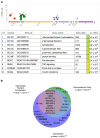
IIIG9 interactors obtained from BioGRID grouped by biological process. (A) Manhattan plot of g: Profiler for IIIG9 interactors: the x-axis shows functional groups by data source (Gene Ontology (GO): Molecular Function, Cellular component, Biological Process; Kyoto Encyclopedia of Genes and Genomes (KEGG); Reactome (REAC); Protein complex (CORUM)) and the y-axis shows the significance in each group. The p-values in the table indicate low (yellow) and high (blue) significance. (B) Venn diagram indicates the subcellular localization of IIIG9 interactors, with respective p-values [42].
Figure 3
Figure 3
IIIG9-interactor proteins organized into semantic groups (UMLS®) of diseases extracted using DisGeNet (A,B) Interactors were experimentally validated (yeast via two-hybrid and affinity capture—MS methods). The semantic groups correspond to concept unique identifiers from the Unified Medical Language system® (UMLS) Metathesaurus ® (version UMLS 2018AA).
Figure 4
Figure 4
Comparison between IIIG9 mRNA expression and protein localization in the adult brain. (A) In situ hybridization analysis in adult mouse brain reported in Allen Brain Atlas for the PPP1R32 gene. (B,C,DD1,FF1) High-magnification image for antisense riboprobe in ependymal cell from lateral and fourth ventricles, neurons from hippocampus, and Purkinje cells from the cerebellum. (B1,EE1,GG1) Immunohistochemical characterization of IIIG9 showing a positive reaction in the cilia and apical membrane of ependymal cells from the dorsal third ventricle and in the soma of hippocampal and Purkinje neurons. (B2). Super-resolution SIM showing IIIG9 dotted pattern (green, arrows) and alpha acetylated tubulin cilia (red) from isolated rat ependymal cells. Sagittal and frontal sections of mouse brain were immunostained with anti-IIIG9 (1:500) and a secondary antibody conjugated with peroxidase. (HL) Immunohistochemical analysis using anti-IIIG9 antibody and anti-IgG labeled with 10 nm gold particles. (HH1) In semi-thin sections, IIIG9 is detected in basal bodies of ependymal cilia (arrow). (IL) In ultra-thin sections, IIIG9 was detected in adherens junctions (arrow), between the ring of nine outer microtubule duplets and the ciliary membrane (arrows), and in the basal bodies (arrow). Magnification in (H): ×100. Scale bars: A: 839 µm; (B1): 50 µm; (E): 1 mm; (G): 60 µm; (J): 1 µm. (BC,DD1,FF1,J1,KL) correspond to digital magnification showing IIIG9 detected in the basal bodies, and between the ring of nine outer microtubule duplets and the membrane of a motile cilia (arrow) (complementary images and results from [6,68]).
Figure 5
Figure 5
IIIG9 expression is restricted to ciliated ependymoma cells of the posterior fossa. (A) Harmony alignment projection of scRNA-seq analysis of 26 pediatric patients with ependymoma (EPN). IIIG9 (PPP1R32) expression is shown in the brown color range and corresponds to the values generated by the adaptive threshold low-rank approximation (ALRA). (BE) IIIG9 expression ALRA range is shown by cluster (B), subtype (C), phase (D), and recurrence (E) The number of cells in each selection is placed in brackets (n = 11200 cells). Seven subgroup clusters of posterior fossa group A (from PFA-sc1 to PFA-sc7), five subgroup clusters of ZFTA-RELA group (from RELA-sc1 to RELA-sc5), one posterior fossa group B (PFB), and one YAP-MAMLD1 group (YAP) are shown. Five PFA subgroup clusters correspond to ciliated EPN cells (CEC), transporter EPN cells (TEC), mesenchymal EPN cells (MEC), and undifferentiated EPN cells 1 and 2 (UEC-1 and EC-2, respectively); cells are in mitosis (mitotic). Uniform manifold approximation and projection (UMAP) was used for classification. The database was generated by Gillen and colleagues [105] and is available in the browsable web resource of the full EPN scRNA-seq dataset at the Pediatric Neuro-oncology Cell Atlas [107].

References

    1. Danielson P.E., Sautkulis L.N., Foye P.E., Hedlund P.B., Carson M.J. A novel mRNA expressed along brain ventricles. Brain Res. Gene Expr. Patterns. 2002;1:83–88. doi: 10.1016/S1567-133X(01)00012-6. - DOI - PubMed
    1. Nucleotide [Internet] Bethesda (MD): National Library of Medicine (US), National Center for Biotechnology Information [1988]. Accession No. NM_145017.3, NM_001170753.2, Homo Sapiens Protein Phosphatase 1 Regulatory Subunit 32 (PPP1R32), mRNA. [(accessed on 10 June 2022)]; Available online: https://www.ncbi.nlm.nih.gov/nuccore/NM_145017.3,NM_001170753.2.
    1. Nucleotide [Internet] Bethesda (MD): National Library of Medicine (US), National Center for Biotechnology Information [1988]. Accession No. NM_133689.2, NM_001368184.1, XM_006527302.5, XM_017318281.1, XM_017318282.1, XM_036161672.1, Mus Musculus Protein Phosphatase 1 Regulatory Subunit 32 (Ppp1r32), mRNA. [(accessed on 10 June 2022)]; Available online: https://www.ncbi.nlm.nih.gov/nuccore/NM_133689.2,NM_001368184.1,XM_00652....
    1. Ivliev A.E., t Hoen P.A., van Roon-Mom W.M., Peters D.J., Sergeeva M.G. Exploring the transcriptome of ciliated cells using in silico dissection of human tissues. PLoS ONE. 2012;7:e35618. doi: 10.1371/journal.pone.0035618. - DOI - PMC - PubMed
    1. Uhlen M., Fagerberg L., Hallstrom B.M., Lindskog C., Oksvold P., Mardinoglu A., Sivertsson A., Kampf C., Sjostedt E., Asplund A., et al. Proteomics. Tissue-based map of the human proteome. Science. 2015;347:1260419. doi: 10.1126/science.1260419. - DOI - PubMed

Publication types