

Identification and Characterization of Elevated Expression of Transferrin and Its Receptor TfR1 in Mouse Models of Depression
- PMID: 36291201
- PMCID: PMC9599150
- DOI: 10.3390/brainsci12101267
Identification and Characterization of Elevated Expression of Transferrin and Its Receptor TfR1 in Mouse Models of Depression
Abstract
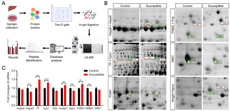
Depression has become one of the severe mental disorders threatening global human health. In this study, we first used the proteomics approach to obtain the differentially expressed proteins in the liver between naive control and chronic social defeat stress (CSDS) induced depressed mice. We have identified the upregulation of iron binding protein transferrin (TF) in the liver, the peripheral blood, and the brain in CSDS-exposed mice. Furthermore, bioinformatics analysis of the Gene Expression Omnibus (GEO) database from various mouse models of depression revealed the significantly upregulated transcripts of TF and its receptor TfR1 in multiple brain regions in depressed mice. We also used the recombinant TF administration via the tail vein to detect its permeability through the blood-brain barrier (BBB). We demonstrated the permeability of peripheral TF into the brain through the BBB. Together, these results identified the elevated expression of TF and its receptor TfR1 in both peripheral liver and the central brain in CSDS-induced depressed mice, and peripheral administration of TF can be transported into the brain through the BBB. Therefore, our data provide a compelling information for understanding the potential role and mechanisms of the cross-talk between the liver and the brain in stress-induced depression.
Keywords: bioinformatics; blood-brain barrier (BBB); chronic social defeat stress (CSDS); depression; mental disorders; proteomics; transferrin (TF); transferrin receptor 1 (TfR1).
Conflict of interest statement
The authors declare no conflict of interest. The funders had no role in the design of the study; in the collection, analyses, or interpretation of data; in the writing of the manuscript; or in the decision to publish the results.
Figures


Similar articles
-
The role of transferrin receptor 1 and 2 in transferrin-bound iron uptake in human hepatoma cells.Am J Physiol Cell Physiol. 2009 Dec;297(6):C1567-75. doi: 10.1152/ajpcell.00649.2008. Epub 2009 Oct 14. Am J Physiol Cell Physiol. 2009. PMID: 19828835
-
Blood-brain barrier transport using a high affinity, brain-selective VNAR antibody targeting transferrin receptor 1.FASEB J. 2021 Feb;35(2):e21172. doi: 10.1096/fj.202001787R. Epub 2020 Nov 25. FASEB J. 2021. PMID: 33241587
-
Effects of cell proliferation on the uptake of transferrin-bound iron by human hepatoma cells.Hepatology. 2003 Oct;38(4):967-77. doi: 10.1053/jhep.2003.50422. Hepatology. 2003. PMID: 14512884
-
Transferrin and transferrin receptors update.Free Radic Biol Med. 2019 Mar;133:46-54. doi: 10.1016/j.freeradbiomed.2018.06.037. Epub 2018 Jun 30. Free Radic Biol Med. 2019. PMID: 29969719 Review.
-
Antibodies Targeting the Transferrin Receptor 1 (TfR1) as Direct Anti-cancer Agents.Front Immunol. 2021 Mar 17;12:607692. doi: 10.3389/fimmu.2021.607692. eCollection 2021. Front Immunol. 2021. PMID: 33815364 Free PMC article. Review.
Cited by
-
Ferroptosis: a new antidepressant pharmacological mechanism.Front Pharmacol. 2024 Jan 8;14:1339057. doi: 10.3389/fphar.2023.1339057. eCollection 2023. Front Pharmacol. 2024. PMID: 38259274 Free PMC article. Review.
-
T cell activation and deficits in T regulatory cells are associated with major depressive disorder and severity of depression.Sci Rep. 2024 May 16;14(1):11177. doi: 10.1038/s41598-024-61865-y. Sci Rep. 2024. PMID: 38750122 Free PMC article.
-
Tyrosine phosphatase SHP2 promoted the progression of CRC via modulating the PI3K/BRD4/TFEB signaling induced ferroptosis.Discov Oncol. 2024 Dec 18;15(1):793. doi: 10.1007/s12672-024-01586-w. Discov Oncol. 2024. PMID: 39692787 Free PMC article.
-
Social defeat stress impairs systemic iron metabolism by activating the hepcidin-ferroportin axis.FASEB Bioadv. 2024 Jul 2;6(8):263-275. doi: 10.1096/fba.2024-00071. eCollection 2024 Aug. FASEB Bioadv. 2024. PMID: 39114446 Free PMC article.
-
Exploring Potential Mechanisms Accounting for Iron Accumulation in the Central Nervous System of Patients with Alzheimer's Disease.Cells. 2024 Apr 16;13(8):689. doi: 10.3390/cells13080689. Cells. 2024. PMID: 38667304 Free PMC article. Review.
References
Grants and funding
LinkOut - more resources
Full Text Sources
Miscellaneous
