. 2022 Sep 23;12(10):1352.
doi: 10.3390/biom12101352.

PHLPP Inhibitor NSC74429 Is Neuroprotective in Rodent Models of Cardiac Arrest and Traumatic Brain Injury

Affiliations

PHLPP Inhibitor NSC74429 Is Neuroprotective in Rodent Models of Cardiac Arrest and Traumatic Brain Injury

Travis C Jackson et al. Biomolecules. .

Abstract

Pleckstrin homology domain and leucine rich repeat protein phosphatase (PHLPP) knockout mice have improved outcomes after a stroke, traumatic brain injury (TBI), and decreased maladaptive vascular remodeling following vascular injury. Thus, small-molecule PHLPP inhibitors have the potential to improve neurological outcomes in a variety of conditions. There is a paucity of data on the efficacy of the known experimental PHLPP inhibitors, and not all may be suited for targeting acute brain injury. Here, we assessed several PHLPP inhibitors not previously explored for neuroprotection (NSC13378, NSC25247, and NSC74429) that had favorable predicted chemistries for targeting the central nervous system (CNS). Neuronal culture studies in staurosporine (apoptosis), glutamate (excitotoxicity), and hydrogen peroxide (necrosis/oxidative stress) revealed that NSC74429 at micromolar concentrations was the most neuroprotective. Subsequent testing in a rat model of asphyxial cardiac arrest, and in a mouse model of severe TBI, showed that serial dosing of 1 mg/kg of NSC74429 over 3 days improved hippocampal survival in both models. Taken together, NSC74429 is neuroprotective across multiple insult mechanisms. Future pharmacokinetic and pharmacodynamic (PK/PD) studies are warranted to optimize dosing, and mechanistic studies are needed to determine the percentage of neuroprotection mediated by PHLPP1/2 inhibition, or potentially from the modulation of PHLPP-independent targets.

Keywords: NSC74429; PHLPP; PHLPP1; PHLPP2; brain; neuroprotection.


Conflict of interest statement

Travis C. Jackson and Patrick M. Kochanek are co-inventors on a pending patent application titled: “Compounds for the treatment of acute organ injury” (USPTO Application No. 16/584,314).

Figures

Figure 1
Figure 1
Physicochemical Properties of PHLPP Inhibitors and the Prediction of Their CNS Activity. (A) An overview of the top-down screening process employed in this study for drug selection. (B) The predicted polar surface aera (PSA) and calculated partition coefficient (CLogP) for each PHLPP inhibitor calculated in Chemicalize (Chemaxon). Compounds highlighted in purple denote the two PHLPP inhibitors that have received the most empirical study/characterization to date. Compounds highlighted in yellow denote unexplored PHLPP inhibitors that have chemistries supportive of BBB penetration. (C) The 2 most studied compounds and the 22 unexplored PHLPP inhibitors organized by known IC50s to block the PP2C domain of PHLPP2. Additionally, the BBB Score (BBB-S) was calculated for each of the compounds and arranged in each subgroup highest to lowest. The red highlighted BBB Scores indicate values < 2 and consistent with non-CNS drugs [6,18]. (D) The chemical structures of 3 unexplored PHLPP inhibitors chosen for testing in neurons is depicted.
Figure 2
Figure 2
Neuroprotective Efficacy of NSC13378, NSC25247, and NSC74429 in Cultured Primary Rat Cortical Neurons. Neurons were injured on day in vitro (DIV) 9 and treated with vehicle or NSC compounds. (A) A scatter plot showing 24 h viability in staurosporine injured neurons treated with NSC13378 or NSC74429. (B) A scatter plot showing 24 h viability in staurosporine injured neurons treated with NSC25247. (C) A scatter plot showing 24 h viability in glutamate injured neurons treated with NSC25247 or NSC74429. (D) A scatter plot showing 24 h viability in hydrogen peroxide injured neurons treated with NSC25247 or NSC74429. All groups include n = 10/group. Data were analyzed by ANOVA and post hoc significance was detected using the Newman-Keuls multiple comparison test. Data were significant at p < 0.05. (*) = p < 0.05, (***) = p < 0.001.
Figure 3
Figure 3
Evaluation of NSC74429 Mediated Neuroprotection in a Rat Model of Cardiac Arrest. (A,B) Normalized CA1 cell count values are shown along with the associated scatter plot. A single CA1 cell count outlier was confirmed by the ROUT method (indicated by the *) and removed prior to statistical analysis of histology. Individual data points highlighted in red in the scatter plot correspond to the rat brains selected as representative images. (CF) Representative images of H&E-stained brains used to assess CA1 cell loss. (G,H) Line graphs (mean + SEM) show changes in overall performance category (OPC) and neurological deficit score (NDS) testing measured in the first 72 h post-injury and at 7 d post-injury. Asterisks indicate significance compared to vehicle-injured rats. Data were significant at p < 0.05. (*) = p < 0.05, (***) = p < 0.001, (****) = p < 0.0001.
Figure 4
Figure 4
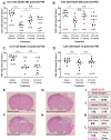
Evaluation of NSC74429 Mediated Neuroprotection in a Mouse Model of TBI. (A,B) Scatter plots of normalized CA1 and CA3 cell counts at 48 h post-injury. (C,D) Scatter plots of normalized CA1 and CA3 cell counts at 7 d post-injury. Individual data points highlighted in red in the scatter plot correspond to the mouse brains selected as representative images in panels E-H. (EH) Representative images of H&E-stained brains used to assess CA1/CA3 cell loss. (IL) Mortality for each group by treatment and study endpoint. Asterisks indicate significance compared to vehicle-injured rats. The red text indicates animals that died. The purple text indicates an animal that survived to the study endpoint but had bleeding in the brain that precluded cell counting. Data were significant at p < 0.05. Not Significant (N.S.), Brain Hemorrhage (B.H.), Controlled Cortical Impact (CCI), Hemorrhagic Shock (HS). (*) = p < 0.05, (**) = p < 0.01, (****) = p < 0.0001.

References

    1. Chen B., Van Winkle J.A., Lyden P.D., Brown J.H., Purcell N.H. PHLPP1 gene deletion protects the brain from ischemic injury. J. Cereb. Blood Flow Metab. 2013;33:196–204. doi: 10.1038/jcbfm.2012.150. - DOI - PMC - PubMed
    1. Jackson T.C., Dixon C.E., Janesko-Feldman K., Vagni V., Kotermanski S.E., Jackson E.K., Kochanek P.M. Acute Physiology and Neurologic Outcomes after Brain Injury in SCOP/PHLPP1 KO Mice. Sci. Rep. 2018;8:7158. doi: 10.1038/s41598-018-25371-2. - DOI - PMC - PubMed
    1. Huang J., Cai C., Zheng T., Wu X., Wang D., Zhang K., Xu B., Yan R., Gong H., Zhang J., et al. Endothelial Scaffolding Protein ENH (Enigma Homolog Protein) Promotes PHLPP2 (Pleckstrin Homology Domain and Leucine-Rich Repeat Protein Phosphatase 2)-Mediated Dephosphorylation of AKT1 and eNOS (Endothelial NO Synthase) Promoting Vascular Remodeling. Arterioscler. Thromb. Vasc. Biol. 2020;40:1705–1721. doi: 10.1161/ATVBAHA.120.314172. - DOI - PubMed
    1. Gao T., Furnari F., Newton A.C. PHLPP: A phosphatase that directly dephosphorylates Akt, promotes apoptosis, and suppresses tumor growth. Mol. Cell. 2005;18:13–24. doi: 10.1016/j.molcel.2005.03.008. - DOI - PubMed
    1. Qiao M., Wang Y., Xu X., Lu J., Dong Y., Tao W., Stein J., Stein G.S., Iglehart J.D., Shi Q., et al. Mst1 is an interacting protein that mediates PHLPPs’ induced apoptosis. Mol. Cell. 2010;38:512–523. doi: 10.1016/j.molcel.2010.03.017. - DOI - PubMed

Publication types