Review
. 2022 Sep 27;12(10):1385.
doi: 10.3390/biom12101385.

Galectin-10 as a Potential Biomarker for Eosinophilic Diseases

Affiliations
Review

Galectin-10 as a Potential Biomarker for Eosinophilic Diseases

Hiroki Tomizawa et al. Biomolecules. .

Abstract

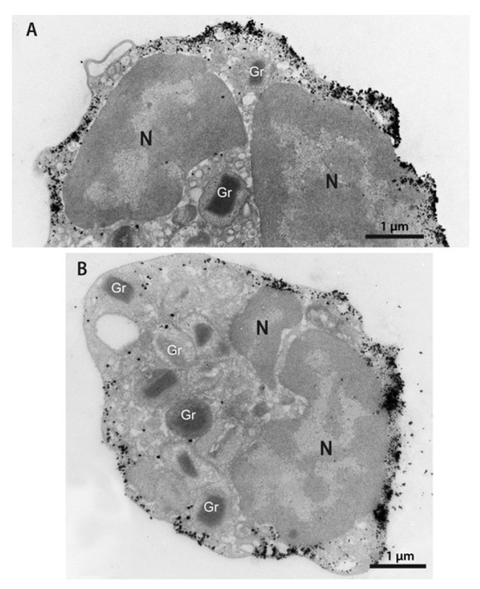
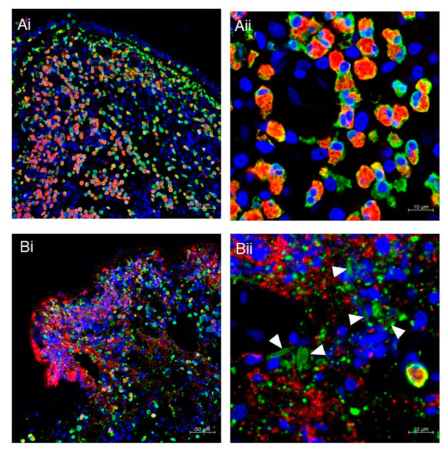
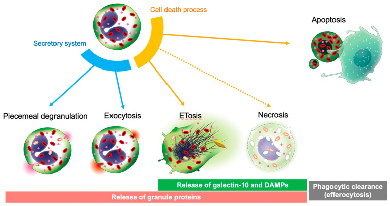
Galectin-10 is a member of the lectin family and one of the most abundant cytoplasmic proteins in human eosinophils. Except for some myeloid leukemia cells, basophils, and minor T cell populations, galectin-10 is exclusively present in eosinophils in the human body. Galectin-10 forms Charcot-Leyden crystals, which are observed in various eosinophilic diseases. Accumulating studies have indicated that galectin-10 acts as a new biomarker for disease activity, diagnosis, and treatment effectiveness in asthma, eosinophilic esophagitis, rhinitis, sinusitis, atopic dermatitis, and eosinophilic granulomatosis with polyangiitis. The extracellular release of galectin-10 is not mediated through conventional secretory processes (piecemeal degranulation or exocytosis), but rather by extracellular trap cell death (ETosis), which is an active cell death program. Eosinophils undergoing ETosis rapidly disintegrate their plasma membranes to release the majority of galectin-10. Therefore, elevated galectin-10 levels in serum and tissue suggest a high degree of eosinophil ETosis. To date, several studies have shown that galectin-10/Charcot-Leyden crystals are more than just markers for eosinophilic inflammation, but play functional roles in immunity. In this review, we focus on the close relationship between eosinophils and galectin-10, highlighting this protein as a potential new biomarker in eosinophilic diseases.

Keywords: Charcot–Leyden crystals; ETosis; eosinophil; eosinophil extracellular trap; galectin-10.


Conflict of interest statement

SU received grants and personal fees from AstraZeneca, personal fees from GlaxoSmithKline and Sanofi, and grants from Novartis and Maruho Co. Ltd.

Figures

Figure 1
Figure 1
(A,B) Distribution of galectin-10 in the eosinophil cytoplasm. Note a robust pool of this protein as show by the deposition of gold particles in the peripheral cytoplasm and in association with the plasma membrane. Secretory granules (Gr) are negative. Isolated human eosinophils were processed for immunonanogold transmission electron microscopy for galectin-10, as previously described [50]. N, nucleus.
Figure 2
Figure 2
(A,B) Immunofluorescence staining of nasal polyps from patients with chronic rhinosinusitis with nasal polyps. Fluorescence images for MBP (red), galectin-10 (green), and DNA (blue) were obtained using a Carl Zeiss LSM780 confocal microscope, as previously described [36]. Intact eosinophils are massively infiltrated in polyps (Ai,Aii). The presence of ETotic eosinophils with extracellular traps (degenerated DNA), cell-free granules (punctate MBP), extracellular vesicles (punctate galectin-10), and CLCs (galectin-10-stained needle-like crystals; arrowheads) can be seen (Bi,Bii). Scale bars: 50 μm for (i) and 10 μm for (ii).
Figure 3
Figure 3
ETosis-mediated galectin-10 release. Live eosinophils secrete granule proteins through piecemeal degranulation and exocytosis. Eosinophils have at least two distinct active cell death pathways of apoptosis and ETosis. Unlike apoptotic cells, which are engulfed and digested by phagocytes (efferocytosis), eosinophils can undergo ETosis in tissue and release their cellular content, including cytoplasmic galectin-10, entire secretory granules, and damage-associated molecular patterns. Therefore, ETosis is also considered a form of eosinophil secretion.

References

    1. Albers F.C., Licskai C., Chanez P., Bratton D.J., Bradford E.S., Yancey S.W., Kwon N., Quirce S. Baseline blood eosinophil count as a predictor of treatment response to the licensed dose of mepolizumab in severe eosinophilic asthma. Respir. Med. 2019;159:105806. doi: 10.1016/j.rmed.2019.105806. - DOI - PubMed
    1. Castro M., Corren J., Pavord I.D., Maspero J., Wenzel S., Rabe K.F., Busse W.W., Ford L., Sher L., Fitzgerald J.M., et al. Dupilumab Efficacy and Safety in Moderate-to-Severe Uncontrolled Asthma. N. Engl. J. Med. 2018;378:2486–2496. doi: 10.1056/NEJMoa1804092. - DOI - PubMed
    1. Humbert M., Taillé C., Mala L., Le Gros V., Just J., Molimard M., STELLAIR Investigators Omalizumab effectiveness in patients with severe allergic asthma according to blood eosinophil count: The STELLAIR study. Eur. Respir. J. 2018;51:1702523. doi: 10.1183/13993003.02523-2017. - DOI - PMC - PubMed
    1. Benson V.S., Hartl S., Barnes N., Galwey N., Van Dyke M.K., Kwon N. Blood eosinophil counts in the general population and airways disease: A comprehensive review and meta-analysis. Eur. Respir. J. 2021;59:2004590. doi: 10.1183/13993003.04590-2020. - DOI - PMC - PubMed
    1. Abidi K., Khoudri I., Belayachi J., Madani N., Zekraoui A., Zeggwagh A.A., Abouqal R. Eosinopenia is a reliable marker of sepsis on admission to medical intensive care units. Crit. Care. 2008;12:R59. doi: 10.1186/cc6883. - DOI - PMC - PubMed

Publication types

MeSH terms

LinkOut - more resources