. 2022 Sep 29;12(10):1395.
doi: 10.3390/biom12101395.

Computational Dissection of the Role of Trp305 in the Regulation of the Death-Associated Protein Kinase-Calmodulin Interaction

Affiliations

Computational Dissection of the Role of Trp305 in the Regulation of the Death-Associated Protein Kinase-Calmodulin Interaction

Yu-Ping Zhu et al. Biomolecules. .

Abstract

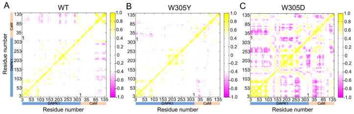
Death-associated protein kinase 1 (DAPK1), as a calcium/calmodulin (CaM) regulated serine/threonine kinase, functions in apoptotic and autophagy pathways and represents an interesting drug target for inflammatory bowel disease and Alzheimer's disease. The crystal structure of the DAPK1 catalytic domain and the autoregulatory domain (ARD) in complex with CaM provides an understanding of CaM-dependent regulation of DAPK1 activity. However, the molecular basis of how distinct Trp305 (W305Y and W305D) mutations in the ARD modulate different DAPK1 activities remains unknown. Here, we performed multiple, μs-length molecular dynamics (MD) simulations of the DAPK1-CaM complex in three different (wild-type, W305Y, and W305D) states. MD simulations showed that the overall structural complex did not change significantly in the wild-type and W305Y systems, but underwent obvious conformational alteration in the W305D system. Dynamical cross-correlation and principal component analyses revealed that the W305D mutation enhanced the anti-correlated motions between the DAPK1 and CaM and sampled a broader distribution of conformational space relative to the wild-type and W305Y systems. Structural and energetical analyses further exhibited that CaM binding was unfavored in response to the W305D mutation, resulting in the decreased binding of CaM to the W305D mutant. Furthermore, the hydrogen bonds and salt bridges responsible for the loss of CaM binding on the interface of the DAPK1-CaM complex were identified in the W305D mutant. This result may provide insights into the key role of Trp305 in the regulation of CaM-mediated DAPK1 activity.

Keywords: binging free energy calculation; calmodulin; death-associated protein kinase 1 (DAPK1); molecular dynamics simulations; principal component analysis.


Conflict of interest statement

The authors declare no conflict of interest.

Figures

Figure 1
Figure 1
(A) A representation of the structural complex of DAPK1–CaM (PDB ID: 2X0G). The catalytic domain (CD) and the autoregulatory domain (ARD) of DAPK1 are colored gray and lemon, respectively. The important basic loop (B-loop), the phosphate-binding loop (P-loop), and the helix αD are colored light blue, lime, and olive, respectively. The N-terminal and the C-terminal CaM are colored pink and light cyan, respectively. Calcium ions are shown by blue spheres. (B) Surface representation of the structural complex of DAPK1–CaM. (C) The hydrophobic network of Trp305 with the CaM residues. Hydrogen bonds are shown by green dotted lines.
Figure 2
Figure 2
The root-mean-square deviation (RMSD) of the protein Cα atoms as a function of simulation time averaged over three independent replicates for the DAPK1–CaM complex (A), the individual DAPK1 (B) and CaM (C) in the wild-type (WT) (red), W305Y (violet), and W305D (yellow) systems. Transparent shading represents standard deviations.
Figure 3
Figure 3
The dynamical cross-correlation matrix (DCCM) plot of the WT (A), W305Y (B), and W305D (C) systems. Anti-correlated/correlated motions with absolute values <0.4 are ignored for clarity and shown in white.
Figure 4
Figure 4
The free energy landscape of the first and second principal components (PC1 vs. PC2) from MD simulations of the WT (A), W305Y (B), and W305D (C) systems. The unit of free-energy values is kcal/mol. The representative DAPK1–CaM structural complex for the WT (A1), W305Y (B1, B2, and B3), and W305D (C1, C2, and C3) are shown.
Figure 5
Figure 5
(AC) The superimposition of the three representative structures (B1, B2, and B3) from the W305Y mutant system on the representative structure A1 from the WT system. (DF). The superimposition of the three representative structures (C1, C2, and C3) from the W305D mutant system on the representative structure A1 from the WT system.
Figure 6
Figure 6
The hydrogen bonds between the interface of DAPK1 and CaM in the WT (A), W305Y (B), and W305D (C) systems. Hydrogen bonds are shown by green dotted lines.
Figure 7
Figure 7
The salt bridges between the interface of DAPK1 and CaM in the WT (A), W305Y (B), and W305D (C) systems. Salt bridges are shown by blue dotted lines.

References

    1. Bialik S., Kimchi A. The death-associated protein kinases: Structure, function, and beyond. Annu. Rev. Biochem. 2006;75:189–210. doi: 10.1146/annurev.biochem.75.103004.142615. - DOI - PubMed
    1. Hupp T.R. Death-associated protein kinase (DAPK) and signal transduction. FEBS J. 2010;277:47. doi: 10.1111/j.1742-4658.2009.07410.x. - DOI - PubMed
    1. Farag A.K., Roh E.J. Death-associated protein kinase (DAPK) family modulators: Current and future therapeutic outcomes. Med. Res. Rev. 2019;39:349–385. doi: 10.1002/med.21518. - DOI - PubMed
    1. Simon B., Huart A.S., Wilmanns M. Molecular mechanisms of protein kinase regulation by calcium/calmodulin. Bioorganic Med. Chem. 2015;23:2749–2760. doi: 10.1016/j.bmc.2015.04.051. - DOI - PubMed
    1. Elbadawy M., Usui T., Yamawaki H., Sasaki K. Novel Functions of Death-Associated Protein Kinases through Mitogen-Activated Protein Kinase-Related Signals. Int. J. Mol. Sci. 2018;19:3031. doi: 10.3390/ijms19103031. - DOI - PMC - PubMed

Publication types

MeSH terms

LinkOut - more resources