. 2022 Oct 7;12(10):1436.
doi: 10.3390/biom12101436.

Looking at the Pathogenesis of the Rabies Lyssavirus Strain Pasteur Vaccins through a Prism of the Disorder-Based Bioinformatics

Affiliations

Looking at the Pathogenesis of the Rabies Lyssavirus Strain Pasteur Vaccins through a Prism of the Disorder-Based Bioinformatics

Surya Dhulipala et al. Biomolecules. .

Abstract

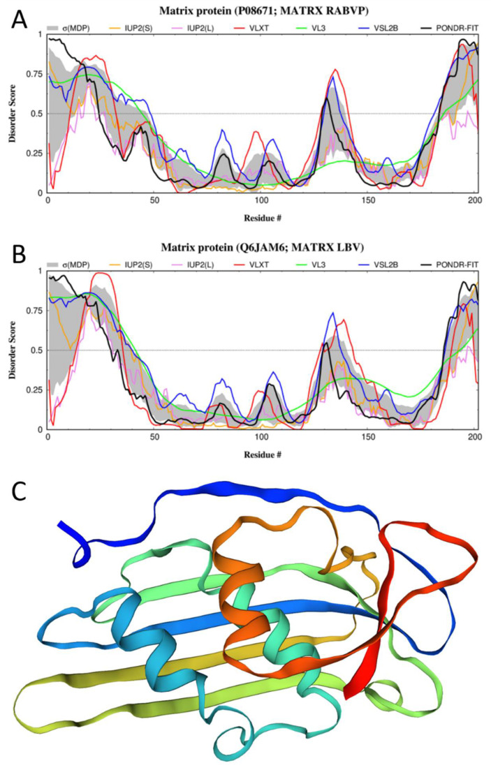
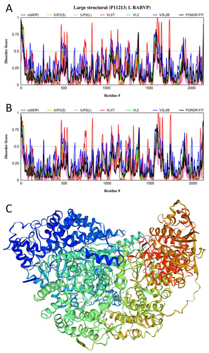
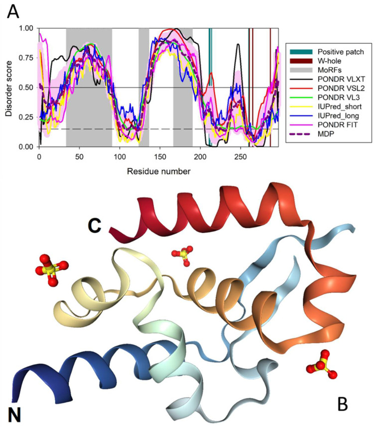
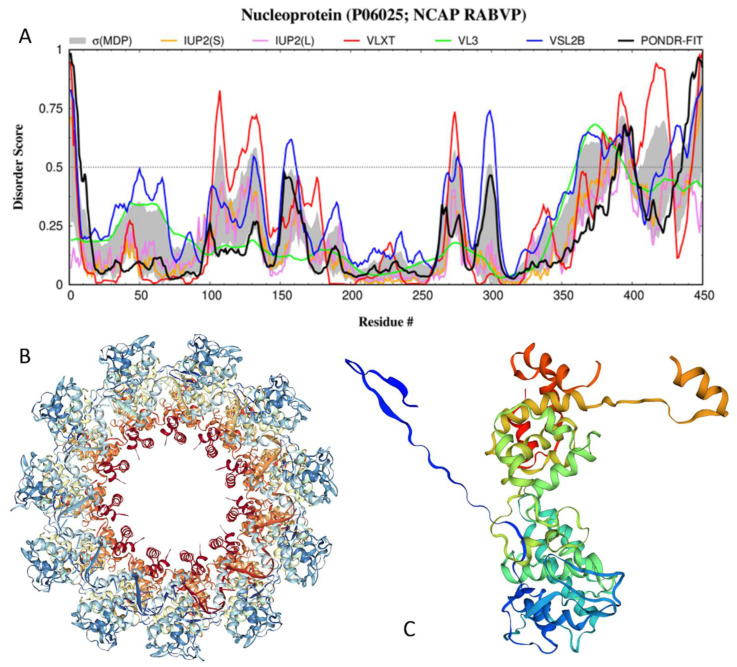
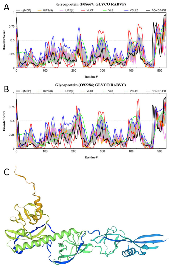
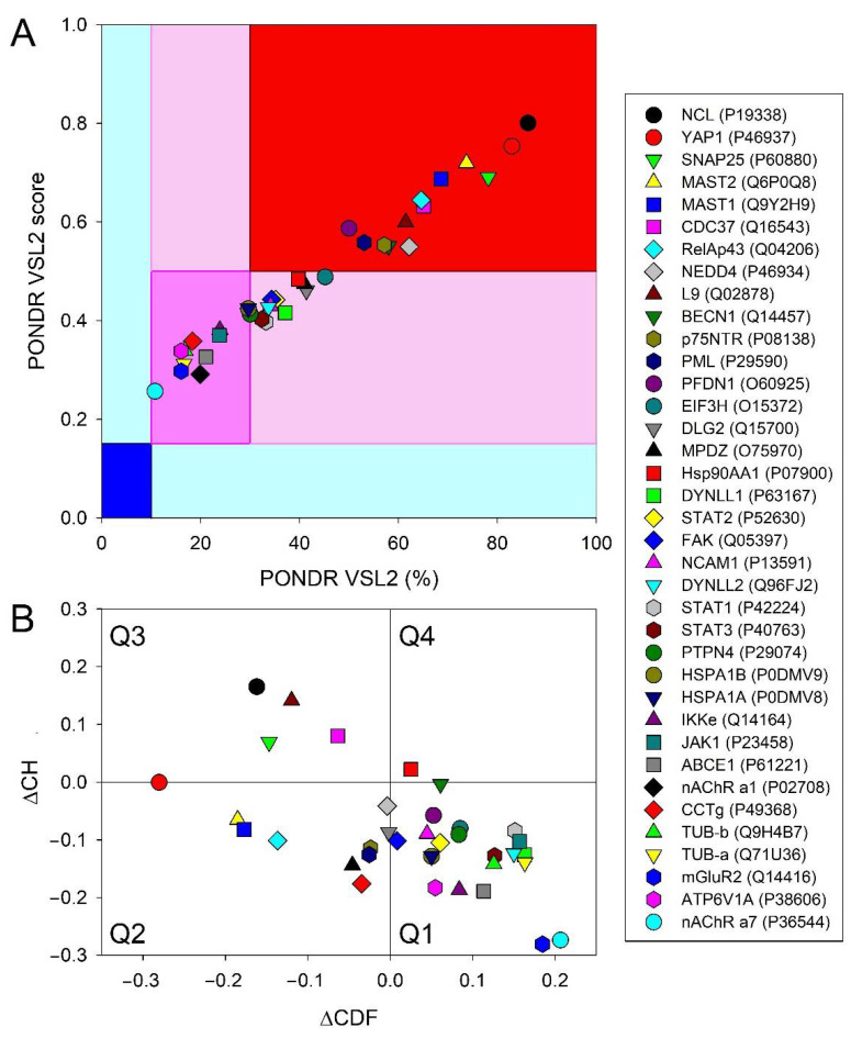
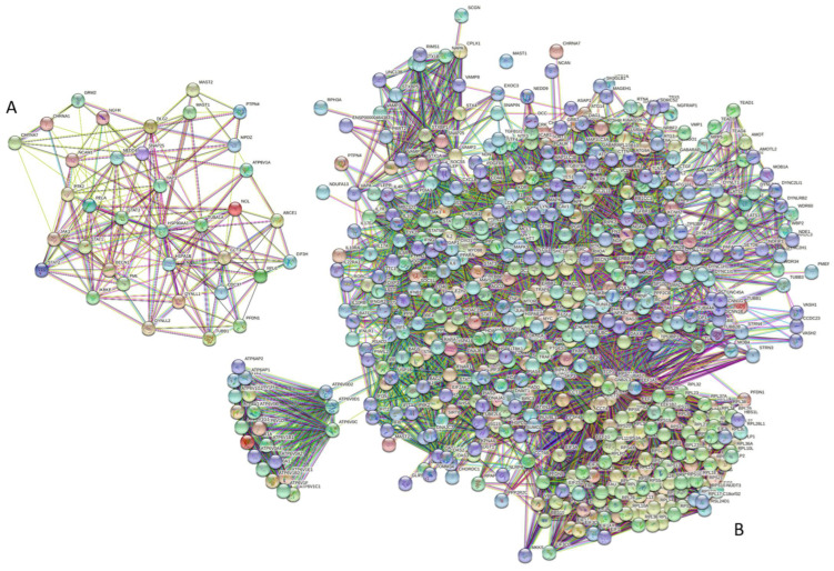
Rabies is a neurological disease that causes between 40,000 and 70,000 deaths every year. Once a rabies patient has become symptomatic, there is no effective treatment for the illness, and in unvaccinated individuals, the case-fatality rate of rabies is close to 100%. French scientists Louis Pasteur and Émile Roux developed the first vaccine for rabies in 1885. If administered before the virus reaches the brain, the modern rabies vaccine imparts long-lasting immunity to the virus and saves more than 250,000 people every year. However, the rabies virus can suppress the host's immune response once it has entered the cells of the brain, making death likely. This study aimed to make use of disorder-based proteomics and bioinformatics to determine the potential impact that intrinsically disordered protein regions (IDPRs) in the proteome of the rabies virus might have on the infectivity and lethality of the disease. This study used the proteome of the Rabies lyssavirus (RABV) strain Pasteur Vaccins (PV), one of the best-understood strains due to its use in the first rabies vaccine, as a model. The data reported in this study are in line with the hypothesis that high levels of intrinsic disorder in the phosphoprotein (P-protein) and nucleoprotein (N-protein) allow them to participate in the creation of Negri bodies and might help this virus to suppress the antiviral immune response in the host cells. Additionally, the study suggests that there could be a link between disorder in the matrix (M) protein and the modulation of viral transcription. The disordered regions in the M-protein might have a possible role in initiating viral budding within the cell. Furthermore, we checked the prevalence of functional disorder in a set of 37 host proteins directly involved in the interaction with the RABV proteins. The hope is that these new insights will aid in the development of treatments for rabies that are effective after infection.

Keywords: intrinsic disorder; intrinsically disordered protein; intrinsically disordered protein region; protein–protein interaction; rabies.


Conflict of interest statement

The authors declare no conflict of interest.

Figures

Figure 3
Figure 3
Structure and disorder in the M-protein from RABV (UniProt ID: P08671). (A) Intrinsic disorder profile (disorder score vs. residues number (residues #)) generated for the M-protein from the RABV strain VP by RIDAO. (B) Intrinsic disorder profile generated for the Lagos bat virus matrix protein (UniProt ID: Q6JAM6) by RIDAO. (C) A structural model for the 30–202 fragment of the RABV M-protein that was created by SWISS-MODEL [97] using the structure of the Lagos bat virus matrix protein (PDB ID: 2W2S; [122]) as a template, which shows sequence identity to the query M-protein of 76.73%.
Figure 6
Figure 6
Structure and disorder in the L-protein from RABV. (A) Intrinsic disorder profile generated for the L-protein from the RABV strain VP (UniProt ID: P11213) by RIDAO. (B) Intrinsic disorder profile generated for the L-protein from the RABV strain SAD B19 by RIDAO (UniProt ID: P16289). (C) A structural model for the L-protein from the RABV strain PV built by SWISS-MODEL [97] using the cryo-EM structure of the large structural protein from the RABV strain SAD B19 (sequence identity: 98.68%) complexed with the fragment of the P-protein (PDB ID: 6UEB, [132]; UniProt ID: P16289) as a template.
Figure 1
Figure 1
Structural features of RABV. In mature RABV, the nucleoprotein, phosphoprotein, and viral polymerase envelop the genomic RNA in a structure known as the ribonucleocapsid (RNP). The matrix protein surrounds the RNP and determines the shape of the virus. The matrix protein also anchors the glycoprotein to the envelope [10] (original source of the image: Philippe Le Mercier, SIB Swiss Institute of Bioinformatics).
Figure 2
Figure 2
Structure and disorder in the RABV P-protein (UniProt ID: P06747). (A) Intrinsic disorder profile generated using data aggregated by the RIDAO platform. The profile also contains disorder/flexibility-related functional annotations and shows three predicted MoRFs (gray-shaded areas), positive patch residues (dark-cyan vertical bars), and W-hole residues (dark-red vertical bars). (B) Crystal structure of the C-terminal region of the RABV P-protein (residues 192–295) (PDB ID: 3OA1).
Figure 4
Figure 4
Structure and disorder in the N-protein from the RABV strain PV (UniProt ID: P06025). (A) Intrinsic disorder profile generated for the M-protein from the RABV strain VP by RIDAO. (B) Crystal structure of the N-protein from the RABV strain ERA (which is 99.11% identical to the N-protein from the RABV strain PV) in complex with RNA (PDB ID: 2GTT; [126]), where the protomers of the N-protein are organized in an undecameric ring. (C) Crystal structure of the N-protein protomer computationally taken out of the undecameric homo-oligomer and demonstrating the presence of two “arms” in the structure (residues 6–28 and 349–414).
Figure 5
Figure 5
Structure and disorder in the G-protein from RABV. (A) Intrinsic disorder profile generated for the G-protein from the RABV strain VP (UniProt ID: P08667) by RIDAO. (B) Intrinsic disorder profile generated for the G-protein from the RABV strain CVS-11 by RIDAO (UniProt ID: O92284). (C) A structural model for the 20–424 fragment of the G-protein from the RABV strain VP generated by SWISS-MODEL [97] using the known structure of the G-protein from the RABV strain CVS-11 (PDB ID: 6LGW [130]; UniProt ID: O92284) as a template, with sequence identity to the query G-protein of 91.48%.
Figure 7
Figure 7
Evaluation of global disorder in 37 human proteins interacting with the RABV proteins. (A) PONDR® VSL2 output for 37 human proteins. The PONDR® VSL2 score is the average disorder score (ADS) for a protein. PONDR® VSL2 (%) is the percent of predicted disordered residues (PPDR). i.e., residues with disorder scores above 0.5. Color blocks indicate regions in which proteins are mostly ordered (blue and light blue), moderately disordered (pink and light pink), or mostly disordered (red). If the two parameters agree, the corresponding part of the background is dark (blue or pink), whereas light blue and light pink reflect areas in which only one of these criteria applies. (B) Charge-hydropathy and cumulative distribution function (CH-CDF) plot. The Y-coordinate is calculated as the distance of the corresponding protein from the boundary in the CH plot. The X-coordinate is calculated as the average distance of the corresponding protein’s CDF curve from the CDF boundary. The quadrant in which the protein is located determines its classification. Q1, protein predicted to be ordered by the CH-plot and CDF. Q2, protein predicted to be ordered by the CH-plot and disordered by the CDF-plot. Q3, protein predicted to be disordered by the CH-plot and CDF. Q4, protein predicted to be disordered by the CH-plot and ordered by CDF.
Figure 8
Figure 8
Intraset and set-based interactivity of human proteins engaged in interactions with the RABV proteins. (A) STRING-generated PPI network with the analyzed set of human proteins. To include all proteins in the network, a low confidence of 0.15 was used as the minimum required interaction score in this case. This network includes 36 proteins linked by 204 interactions. The resulting average node degree of this network is 11.3, and its average local clustering coefficient (which defines how close its neighbors are to being a complete clique; the local clustering coefficient is equal to 1 if every neighbor connected to a given node Ni is also connected to every other node within the neighborhood, and it is equal to 0 if no node that is connected to a given node Ni connects to any other node that is connected to Ni) is 0.578. Since the expected number of edges in a network of the same size for proteins randomly selected from the human proteome is 128, this network is characterized by a PPI enrichment p-value of 4.7 × 10⁻¹⁰. (B) The STRING-generated PPI network centered on human proteins interacting with the RABV proteins. Note that the number of interactors in STRING is limited to 500. This network, generated with a high confidence score of 0.7, includes 536 proteins connected by 11,358 interactions. The average node degree and average local clustering coefficient of this PPI are 42.4 and 0.631, respectively; its PPI enrichment p-value is <10−16.
Figure 9
Figure 9
Disordered interactome of the RABV N-, L-, P-, M-, and G-proteins. Proteins are colored based on their PPIDR values, with highly and moderately disordered proteins being shown by red and pink colors, respectively. Note that none of the proteins in this diagram is classified as mostly ordered (there are no proteins colored in blue).

Similar articles

Cited by

References

    1. Rupprecht C.E. Rhabdoviruses: Rabies Virus. In: Baron S., editor. Medical Microbiology. 4th ed. University of Texas Medical Branch; Galveston, TX, USA: 1996. - PubMed
    1. Pieracci E.G., Pearson C.M., Wallace R.M., Blanton J.D., Whitehouse E.R., Ma X., Stauffer K., Chipman R.B., Olson V. Vital Signs: Trends in Human Rabies Deaths and Exposures—United States, 1938–2018. MMWR Morb. Mortal. Wkly. Rep. 2019;68:524–528. doi: 10.15585/mmwr.mm6823e1. - DOI - PMC - PubMed
    1. Dietzschold B., Li J., Faber M., Schnell M. Concepts in the pathogenesis of rabies. Futur. Virol. 2008;3:481–490. doi: 10.2217/17460794.3.5.481. - DOI - PMC - PubMed
    1. Gluska S., Zahavi E.E., Chein M., Gradus T., Bauer A., Finke S., Perlson E. Rabies Virus Hijacks and Accelerates the p75NTR Retrograde Axonal Transport Machinery. PLoS Pathog. 2014;10:e1004348. doi: 10.1371/journal.ppat.1004348. - DOI - PMC - PubMed
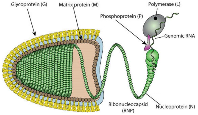
    1. ViralZone. Lyssavirus. [(accessed on 20 July 2020)]. Available online: https://viralzone.expasy.org/resources/Rhabdoviridae_virion.jpg.

MeSH terms