. 2022 Oct 14;12(10):1480.
doi: 10.3390/biom12101480.

Quantifying Coexistence Concentrations in Multi-Component Phase-Separating Systems Using Analytical HPLC

Affiliations

Quantifying Coexistence Concentrations in Multi-Component Phase-Separating Systems Using Analytical HPLC

Anne Bremer et al. Biomolecules. .

Abstract

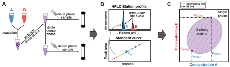
Over the last decade, evidence has accumulated to suggest that numerous instances of cellular compartmentalization can be explained by the phenomenon of phase separation. This is a process by which a macromolecular solution separates spontaneously into dense and dilute coexisting phases. Semi-quantitative, in vitro approaches for measuring phase boundaries have proven very useful in determining some key features of biomolecular condensates, but these methods often lack the precision necessary for generating quantitative models. Therefore, there is a clear need for techniques that allow quantitation of coexisting dilute and dense phase concentrations of phase-separating biomolecules, especially in systems with more than one type of macromolecule. Here, we report the design and deployment of analytical High-Performance Liquid Chromatography (HPLC) for in vitro separation and quantification of distinct biomolecules that allows us to measure dilute and dense phase concentrations needed to reconstruct coexistence curves in multicomponent mixtures. This approach is label-free, detects lower amounts of material than is accessible with classic UV-spectrophotometers, is applicable to a broad range of macromolecules of interest, is a semi-high-throughput technique, and if needed, the macromolecules can be recovered for further use. The approach promises to provide quantitative insights into the balance of homotypic and heterotypic interactions in multicomponent phase-separating systems.

Keywords: biomolecular condensates; coexistence line; phase separation.


Conflict of interest statement

Tanja Mittag is a member of the Scientific Advisory Board of Faze Medicines. Rohit Pappu is a member of the Scientific Advisory Board of Dewpoint Therapeutics. The work reported here was not influenced by these affiliations.

Figures

Figure 1
Figure 1
Schematic overview of the workflow used in this study to reconstruct phase boundaries via analytical HPLC. (A) Biomolecules A and B undergo phase separation when present at suitable concentrations and molar ratios. The sample is incubated for equilibration. Separation of the dense and dilute phases is achieved via centrifugation. Known volumes of dilute and dense phase are each separately injected onto the HPLC column and eluted with an appropriate method. (B) HPLC elution profile for a sample containing biomolecules A and B. Peaks are integrated and the concentrations of biomolecules A and B are determined using a standard curve. (C) The coexistence line and tie lines of biomolecules A and B can be reconstructed from the dilute and dense phase concentrations extracted from the elution profiles. The tie line connects the coexisting dilute and dense phase concentrations. A tie line or tie simplex is defined by its slope, and it identifies the components whose concentrations need to be constrained relative to one another to yield the concentrations of the coexisting phases.
Figure 2
Figure 2
Determination of csat and cdense by analytical HPLC. (A) The standard curve for A1-LCD was determined by plotting the area under the HPLC elution peak (IA1-LCD) from injections of different amounts of A1-LCD, nA1-LCD in nmoles. (B) Comparison of csat and cdense values obtained via measurements using HPLC vs values from UV absorption measurements by spectrophotometer for A1-LCD as a function of temperature [36]. The dashed line represents a fit of the Flory Huggins equation to the spectrophotometer data. The shaded area thus represents the 2-phase regime determined by the coexistence concentrations and other extant data [35,36].
Figure 3
Figure 3
Determination of dilute and dense phase concentrations for multi-component systems. (A) Dilute phase concentrations of Gcn4 in the presence of increasing concentrations of PEG8000. Individual measurements are shown in grey, with the average shown in green. At least three replicates per sample were measured and error bars represent the standard deviation. (B) HPLC chromatogram showing the elution profile of samples of the dilute and dense phase for the A1-LCD/FUS-PLD mixture. The dense phase sample was diluted prior to injection. For a comparison of absorbance at 230 and 280 nm, please see Figure S2. (C) csat and cdense for A1-LCD and FUS-PLD for the case of their homotypic or heterotypic phase separation. (D) Data points from C are shown in a 2D phase diagram. The tie line between coexisting dilute and dense phase concentrations in the heterotypic system is shown as dashed line. The 2-phase regime is approximated as shaded area as expected from few presented data points.

References

    1. Shin Y., Brangwynne C.P. Liquid phase condensation in cell physiology and disease. Science. 2017;357 doi: 10.1126/science.aaf4382. - DOI - PubMed
    1. Banani S.F., Lee H.O., Hyman A.A., Rosen M.K. Biomolecular condensates: Organizers of cellular biochemistry. Nat. Rev. Mol. Cell Biol. 2017;18:285–298. - PMC - PubMed
    1. Holehouse A.S., Pappu R.V. Functional Implications of Intracellular Phase Transitions. Biochemistry. 2018;57:2415–2423. doi: 10.1021/acs.biochem.7b01136. - DOI - PMC - PubMed
    1. Alberti S., Gladfelter A., Mittag T. Considerations and Challenges in Studying Liquid-Liquid Phase Separation and Biomolecular Condensates. Cell. 2019;176:419–434. doi: 10.1016/j.cell.2018.12.035. - DOI - PMC - PubMed
    1. Mittag T., Pappu R.V. A conceptual framework for understanding phase separation and addressing open questions and challenges. Mol. Cell. 2022;82:2201–2214. doi: 10.1016/j.molcel.2022.05.018. - DOI - PMC - PubMed

Publication types

MeSH terms

Substances

LinkOut - more resources