. 2022 Oct 16;12(10):1493.
doi: 10.3390/biom12101493.

Gene Delivery of Manf to Beta-Cells of the Pancreatic Islets Protects NOD Mice from Type 1 Diabetes Development

Affiliations

Gene Delivery of Manf to Beta-Cells of the Pancreatic Islets Protects NOD Mice from Type 1 Diabetes Development

Kailash Singh et al. Biomolecules. .

Abstract

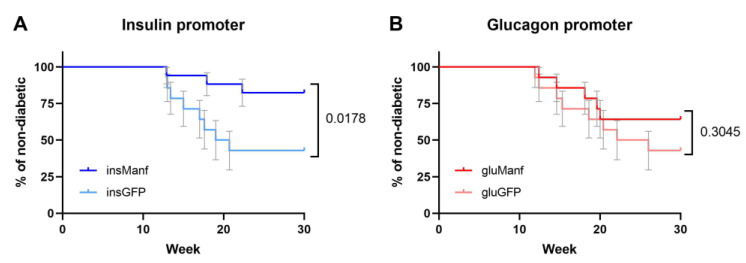
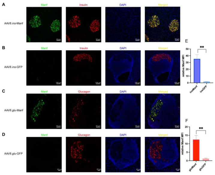
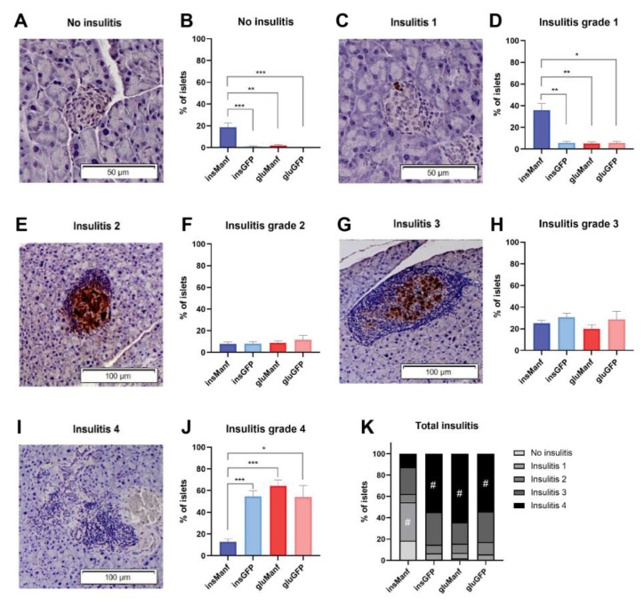
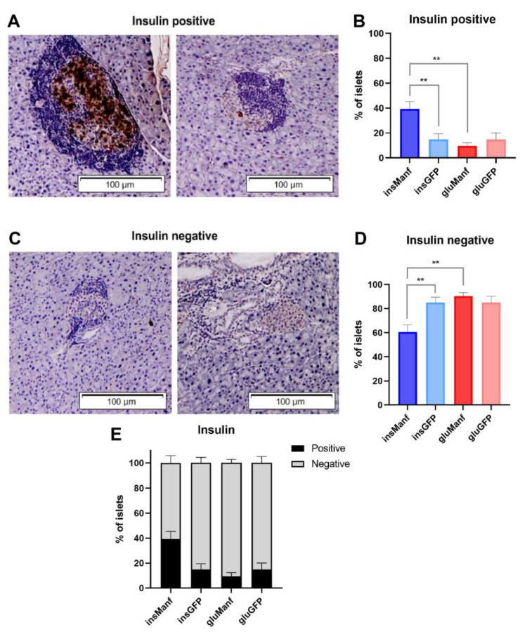
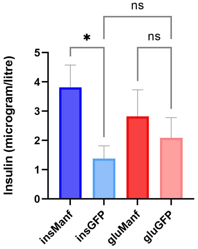
In type 1 diabetes, dysfunctional glucose regulation occurs due to the death of insulin-producing beta-cells in the pancreatic islets. Initiation of this process is caused by the inheritance of an adaptive immune system that is predisposed to responding to beta-cell antigens, most notably to insulin itself, coupled with unknown environmental insults priming the autoimmune reaction. While autoimmunity is a primary driver in beta-cell death, there is growing evidence that cellular stress participates in the loss of beta-cells. In the beta-cell fragility model, partial loss of islet mass requires compensatory upregulation of insulin production in the remaining islets, driving a cellular stress capable of triggering apoptosis in the remaining cells. The Glis3-Manf axis has been identified as being pivotal to the relative fragility or robustness of stressed islets, potentially operating in both type 1 and type 2 diabetes. Here, we have used an AAV-based gene delivery system to enhance the expression of the anti-apoptotic protein Manf in the beta-cells of NOD mice. Gene delivery substantially lowered the rate of diabetes development in treated mice. Manf-treated mice demonstrated minimal insulitis and superior preservation of insulin production. Our results demonstrating the therapeutic potential of Manf delivery to enhance beta-cell robustness and avert clinical diabetes.

Keywords: AAV; Manf; NOD mice; beta-cells; gene delivery; type 1 diabetes.


Conflict of interest statement

This work is the subject of active commercialization efforts by the Babraham Institute and VIB, which may result in financial return to authors. The funders had no role in the design of the study; in the collection, analyses, or interpretation of data; in the writing of the manuscript; or in the decision to publish the results.

Figures

Figure 1
Figure 1
AAV8.ins-Manf prevented diabetes development in NOD mice. The percentage of non-diabetic NOD mice was controlled after the end of treatment at 30 weeks of age. (A) The AAV8.ins-Manf group (n = 17) and the AAV8.ins-GFP group (n = 14) had 82.4% and 42.8% non-diabetic mice, respectively (p-value of 0.0178). (B) The comparison between two groups with glucagon promoters, AAV8.glu-Manf (n = 14) and AAV8.glu-GFP (n = 14), had a p-value of 0.3045, which show no statistically significant difference. For comparison a Log-rank test was used.
Figure 2
Figure 2
Islets of AAV8.ins-Manf- or AAV8.glu-Manf-treated mice express Manf. (AD) AAV8.ins-Manf-, AAV8.ins-GFP-, AAV8.glu-Manf-, and AAV8.glu-GFP-treated mice were stained with antibodies: Manf (green), insulin (red), or glucagon (red) and DAPI (blue). (E,F) mean fluorescence intensity (MFI) of Manf in islets of AAV8.ins-Manf-, AAV8.ins-GFP-, AAV8.glu-Manf-, and AAV8.glu-GFP-treated mice. Three to four slides from each mouse (in total 4 mice per group) were stained with antibodies: Manf, insulin, or glucagon and DAPI. Confocal images were captured and analysed using ImageJ software for determining MFI. Results are presented in means ± SEM (n = 4 per group). Unpaired t-tests were performed for comparison, ** denote p < 0.05.
Figure 3
Figure 3
Beta-cell-specific gene delivery of Manf prevented islets from severe insulitis. Light microscope pictures of islets representing no insulitis (A), insulitis grade 1 (C), insulitis grade 2 (E), insulitis grade 3 (G), and insulitis grade 4 (I), are demonstrated as well as graphs of each insulitis grading, comparing the mean percentages of islets in each group (B,D,F,H,J). (K) Total insulitis. Results are presented in means ± SEM (n = 14–17/group). Kruskal-Wallis test, followed by the Dunn’s test was performed for multiple comparisons for finding the significant differences. *, ** and *** denote p < 0.05, p < 0.01 and p < 0.001, respectively.
Figure 4
Figure 4
Beta-cell-specific gene delivery of Manf increased the content of insulin in islets of NOD mice. Light microscope pictures of NOD mice tissue sections immunohistochemically stained for insulin (A); a representative image of islets of AAV8.ins-Manf treated mice and (C); a representative image of islets of AAV8.ins-GFP treated mice) and graphs over the mean percentage of insulin-positive or insulin-negative islets in each group (B,D). (A) Two microscope pictures representing insulin-positive islets, where brown colour indicates insulin. (B) The percentages of insulin-positive islets in treated mice. (C) Two microscope pictures representing insulin-negative islets. (D) The percentages of insulin-negative islets in treated mice. (E) Percentages of insulin-positive and insulin-negative cells in total. Results are presented in means ± SEM (n = 14–17/group). Kruskal–Wallis test followed by Dunn’s test was performed for multiple comparisons. ** denote p < 0.01.
Figure 5
Figure 5
Beta-cell-specific gene delivery of Manf increased the serum levels of insulin in islets of NOD mice. Serum samples were analysed using Ultra-Sensitive Insulin ELISA kit. Results are presented in means ± SEM (n = 11–15/group). Kruskal–Wallis test followed by Dunn’s test was performed for multiple comparisons. * denotes p < 0.05.

Similar articles

Cited by

References

    1. Roizen J.D., Bradfield J.P., Hakonarson H. Progress in understanding type 1 diabetes through its genetic overlap with other autoimmune diseases. Curr. Diab. Rep. 2015;15:102. doi: 10.1007/s11892-015-0668-4. - DOI - PMC - PubMed
    1. Bonifacio E., Mathieu C., Nepom G.T., Ziegler A.G., Anhalt H., Haller M.J., Harrison L.C., Hebrok M., Kushner J.A., Norris J.M., et al. Rebranding asymptomatic type 1 diabetes: The case for autoimmune beta cell disorder as a pathological and diagnostic entity. Diabetologia. 2017;60:35–38. doi: 10.1007/s00125-016-4144-8. - DOI - PMC - PubMed
    1. Heianza Y., Arase Y., Fujihara K., Tsuji H., Saito K., Hsieh S.D., Kodama S., Shimano H., Yamada N., Hara S., et al. Screening for pre-diabetes to predict future diabetes using various cut-off points for HbA(1c) and impaired fasting glucose: The Toranomon Hospital Health Management Center Study 4 (TOPICS 4) Diabet. Med. 2012;29:e279–e285. doi: 10.1111/j.1464-5491.2012.03686.x. - DOI - PubMed
    1. Cnop M., Welsh N., Jonas J.C., Jorns A., Lenzen S., Eizirik D.L. Mechanisms of pancreatic beta-cell death in type 1 and type 2 diabetes: Many differences, few similarities. Diabetes. 2005;54((Suppl. 02)):S97–S107. doi: 10.2337/diabetes.54.suppl_2.S97. - DOI - PubMed
    1. Butler A.E., Janson J., Bonner-Weir S., Ritzel R., Rizza R.A., Butler P.C. Beta-cell deficit and increased beta-cell apoptosis in humans with type 2 diabetes. Diabetes. 2003;52:102–110. doi: 10.2337/diabetes.52.1.102. - DOI - PubMed

Publication types