

Mechanisms of Peritoneal Mesothelial Cells in Peritoneal Adhesion
- PMID: 36291710
- PMCID: PMC9599397
- DOI: 10.3390/biom12101498
Mechanisms of Peritoneal Mesothelial Cells in Peritoneal Adhesion
Abstract
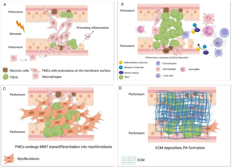
A peritoneal adhesion (PA) is a fibrotic tissue connecting the abdominal or visceral organs to the peritoneum. The formation of PAs can induce a variety of clinical diseases. However, there is currently no effective strategy for the prevention and treatment of PAs. Damage to peritoneal mesothelial cells (PMCs) is believed to cause PAs by promoting inflammation, fibrin deposition, and fibrosis formation. In the early stages of PA formation, PMCs undergo mesothelial-mesenchymal transition and have the ability to produce an extracellular matrix. The PMCs may transdifferentiate into myofibroblasts and accelerate the formation of PAs. Therefore, the aim of this review was to understand the mechanism of action of PMCs in PAs, and to offer a theoretical foundation for the treatment and prevention of PAs.
Keywords: fibrosis; inflammation; mesothelial–mesenchymal transition; peritoneal adhesions; peritoneal mesothelial cells.
Conflict of interest statement
The authors declare no conflict of interest.
Figures
References
-
- Tsai J.M., Sinha R., Seita J., Fernhoff N., Christ S., Koopmans T., Krampitz G.W., McKenna K.M., Xing L., Sandholzer M., et al. Surgical adhesions in mice are derived from mesothelial cells and can be targeted by antibodies against mesothelial markers. Sci. Transl. Med. 2018;10:eaan6735. doi: 10.1126/scitranslmed.aan6735. - DOI - PubMed
-
- Kalra A., Wehrle C.J., Tuma F. Anatomy, Abdomen and Pelvis, Peritoneum. StatPearls Publishing LLC.; Treasure Island, FL, USA: 2022. - PubMed
Publication types
MeSH terms
Substances
LinkOut - more resources
Full Text Sources
