Review
. 2022 Oct 13;14(20):5009.
doi: 10.3390/cancers14205009.

The Tumor Microenvironment of Medulloblastoma: An Intricate Multicellular Network with Therapeutic Potential

Affiliations
Review

The Tumor Microenvironment of Medulloblastoma: An Intricate Multicellular Network with Therapeutic Potential

Niek F H N van Bree et al. Cancers (Basel). .

Abstract

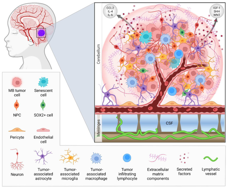
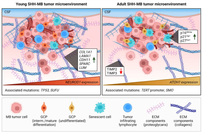
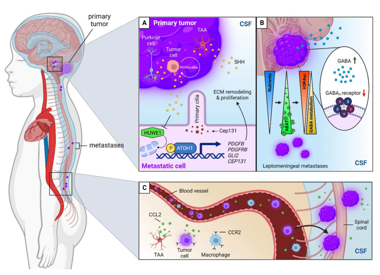
Medulloblastoma (MB) is a heterogeneous disease in which survival is highly affected by the underlying subgroup-specific characteristics. Although the current treatment modalities have increased the overall survival rates of MB up to 70-80%, MB remains a major cause of cancer-related mortality among children. This indicates that novel therapeutic approaches against MB are needed. New promising treatment options comprise the targeting of cells and components of the tumor microenvironment (TME). The TME of MB consists of an intricate multicellular network of tumor cells, progenitor cells, astrocytes, neurons, supporting stromal cells, microglia, immune cells, extracellular matrix components, and vasculature systems. In this review, we will discuss all the different components of the MB TME and their role in MB initiation, progression, metastasis, and relapse. Additionally, we briefly introduce the effect that age plays on the TME of brain malignancies and discuss the MB subgroup-specific differences in TME components and how all of these variations could affect the progression of MB. Finally, we highlight the TME-directed treatments, in which we will focus on therapies that are being evaluated in clinical trials.

Keywords: age-associated differences; brain tumor vasculature; extracellular matrix; immune cells; leptomeningeal dissemination; medulloblastoma; tumor microenvironment.


Conflict of interest statement

The authors declare no conflict of interest.

Figures

Figure 1
Figure 1
The tumor microenvironment of human medulloblastoma (MB). A MB tumor consists of an intricate multicellular network with extracellular matrix (ECM) components. These tumors are thought to arise from embryonic progenitor cells such as neural progenitor cells (NPCs). Senescent cells and SOX2+ cells form modes through which recurrence or relapse can occur. The blood–brain barrier (BBB) is an important part of the tumor microenvironment (TME) of MB. It consists of pericytes and astrocyte foot processes that surround the blood vessels, which are composed of a basement membrane and specialized endothelial cells. In most MBs the BBB is intact, functioning as a highly selective border. However, this integrity is influenced by the MB subgroup. The defining pattern of metastasis for MB is leptomeningeal dissemination, in which tumor cells can spread to the meninges through the cerebrospinal fluid (CSF). The lymphatic vasculature in the brain has only been recently discovered [19,20]. The role of lymphatic vessels in MB has, therefore, not been studied yet. However, this system might prove to be an important asset of the TME of MB. Furthermore, the TME of MB consists of unique brain-resident cell types such as microglia [21], astrocytes [22], and neurons [12], each associated with MB tumor progression in distinctive ways. MB has a low occurrence of tumor-infiltrating lymphocytes (T and B cells) and other immune cells [23]. Most abundant are the tissue-resident microglia and tumor-associated macrophages. All cells within the TME are responsible for the production of secreted factors such as growth factors, cytokines, and chemokines (e.g., IL-4, IL-6, CCL2, IGF-1, SHH, WNT) that stimulate tumor progression [22,24,25]. Support for this multicellular tumor network is provided by ECM components. MBs are highly enriched in glycoproteins (e.g., laminin and vitronectin) and proteoglycan-degrading enzymes (e.g., heparanase), causing dysregulation of the brain’s ECM composition.
Figure 2
Figure 2
Age-associated differences in the tumor microenvironments of young and adult human SHH-driven medulloblastoma (MB). Age affects both the cellular and extracellular matrix (ECM) compositions of the MB tumor microenvironment (TME). Pediatric SHH-MB tumors contain relatively more tumor-infiltrating lymphocytes [23], whereas dormant senescent cells are more associated with the development of SHH-MB tumors in adults [33,44]. Preneoplastic MB lesions in adults show increased expression of senescence markers p16INK4a, p21Cip1, and p27Kip1. Additionally, young and adult SHH-MB tumors display varying orders of granule cell precursor (GCP) differentiation. NEUROD1+ GCPs in pediatric SHH-MB is a sign of an intermediate to mature differentiation state, while ATOH1 expression marks an undifferentiated state of GCPs in adult SHH-MB. Furthermore, ECM components, such as proteoglycans and collagens, are enriched in pediatric SHH-MB tumors. This has been observed both by an increase in ECM-related genes (e.g., COL1A1, LAMA1, CDH11, SPARC, LUM) in pediatric SHH-MB, as well as a decrease in ECM enzymatic inhibitors (TIMP2 and TIMP3) in adult SHH-MB. Lastly, TP53 and SUFU mutations are more associated with pediatric SHH-MB, whereas TERT promoter and SMO mutations are mainly found in adult SHH-MB tumors. CSF = cerebrospinal fluid. Green arrow = increased expression; red arrow = decreased expression.
Figure 3
Figure 3
TME-associated mechanisms of leptomeningeal dissemination (LMD) in medulloblastoma (MB). (A) Aberrant SHH signaling and ATOH1 expression promote LMD in SHH-MB. SHH ligand is produced by Purkinje cells, MB tumor cells, and tumor-associated astrocytes (TAA) and stimulates the continuous proliferation of tumor cells through the activation of GLI and ATOH1 transcription factors. ATOH1 can control the formation of primary cilia by regulating the expression of Cep131, allowing for SHH-triggered proliferation, and also influences extracellular matrix (ECM) remodeling by activating ECM remodeling genes such as PDGFB and PDGFRB enabling metastatic spread. Additionally, SHH prevents the degradation of ATOH1 by blocking HUWE1. (B) Metastatic MB cells use GABA transaminase (ABAT) to metabolize GABA to survive in the nutrient-deficient cerebrospinal fluid (CSF). The metastatic cells adapt mature GABAergic neuronal characteristics such as H3K4ac histone deacetylation and GABA metabolism. Furthermore, GABAA receptor activity is decreased in the primary MB tumor and leptomeningeal metastases, yielding more free circulating GABA molecules in the body. Green arrow = increased availability; red arrow = decreased activity. (C) LMD can also occur via a hematogenous route via the activation of the CCL2–CCR2 axis. CCL2 is, among others, secreted by TAAs and tumor cells. Both tumor cells and (tumor-associated) macrophages contain CCR2 receptors for CCL2 signaling. The CCL2 levels are increased in the CSF of MB patients.

Similar articles

Cited by

References

    1. Majd N., Penas-Prado M. Updates on Management of Adult Medulloblastoma. Curr Treat. Options Oncol. 2019;20:64. doi: 10.1007/s11864-019-0663-0. - DOI - PubMed
    1. Juraschka K., Taylor M.D. Medulloblastoma in the age of molecular subgroups: A review. J. Neurosurg. Pediatr. 2019;24:353–363. doi: 10.3171/2019.5.PEDS18381. - DOI - PubMed
    1. Cavalli F.M.G., Remke M., Rampasek L., Peacock J., Shih D.J.H., Luu B., Garzia L., Torchia J., Nor C., Morrissy A.S., et al. Intertumoral Heterogeneity within Medulloblastoma Subgroups. Cancer Cell. 2017;31:737–754 e736. doi: 10.1016/j.ccell.2017.05.005. - DOI - PMC - PubMed
    1. Menyhart O., Giangaspero F., Gyorffy B. Molecular markers and potential therapeutic targets in non-WNT/non-SHH (group 3 and group 4) medulloblastomas. J. Hematol. Oncol. 2019;12:29. doi: 10.1186/s13045-019-0712-y. - DOI - PMC - PubMed
    1. Riemondy K.A., Venkataraman S., Willard N., Nellan A., Sanford B., Griesinger A.M., Amani V., Mitra S., Hankinson T.C., Handler M.H., et al. Neoplastic and immune single-cell transcriptomics define subgroup-specific intra-tumoral heterogeneity of childhood medulloblastoma. Neuro. Oncol. 2022;24:273–286. doi: 10.1093/neuonc/noab135. - DOI - PMC - PubMed

LinkOut - more resources