. 2022 Oct 14;14(20):5028.
doi: 10.3390/cancers14205028.

Genomic Instability in Cerebrospinal Fluid Cell-Free DNA Predicts Poor Prognosis in Solid Tumor Patients with Meningeal Metastasis

Affiliations

Genomic Instability in Cerebrospinal Fluid Cell-Free DNA Predicts Poor Prognosis in Solid Tumor Patients with Meningeal Metastasis

Peng Wang et al. Cancers (Basel). .

Abstract

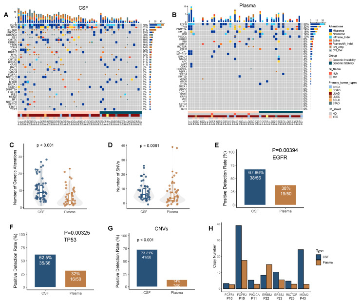
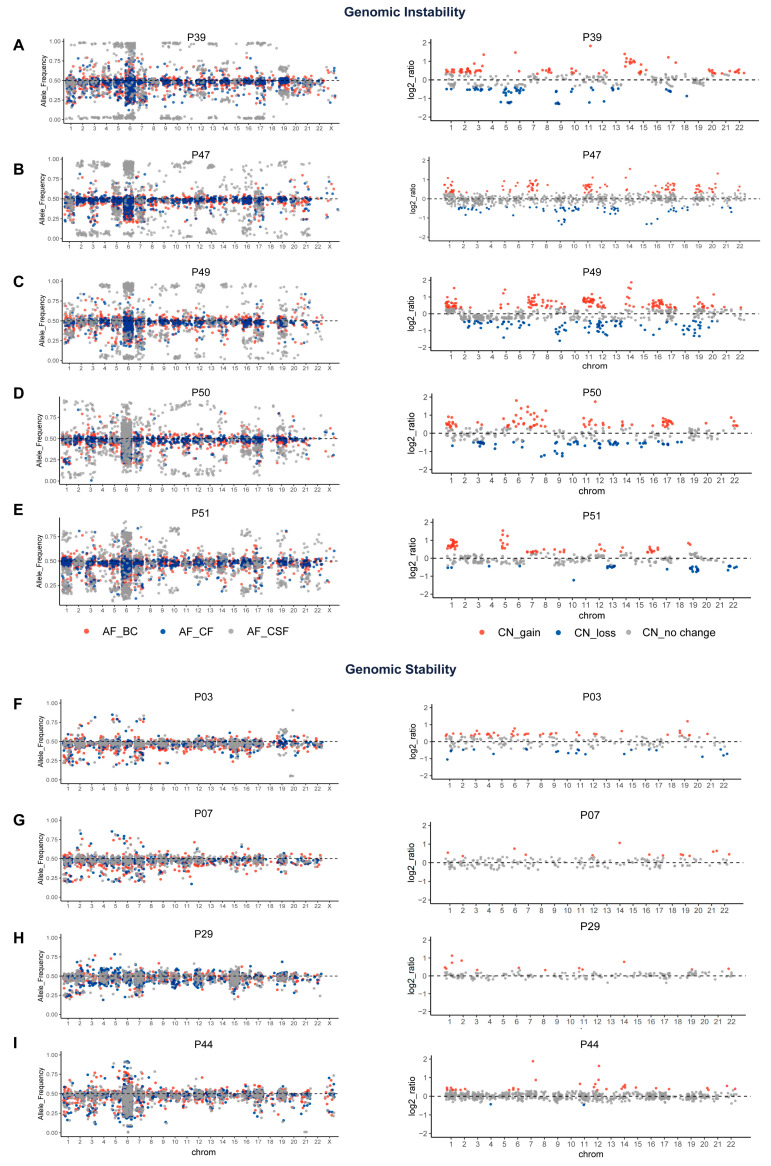
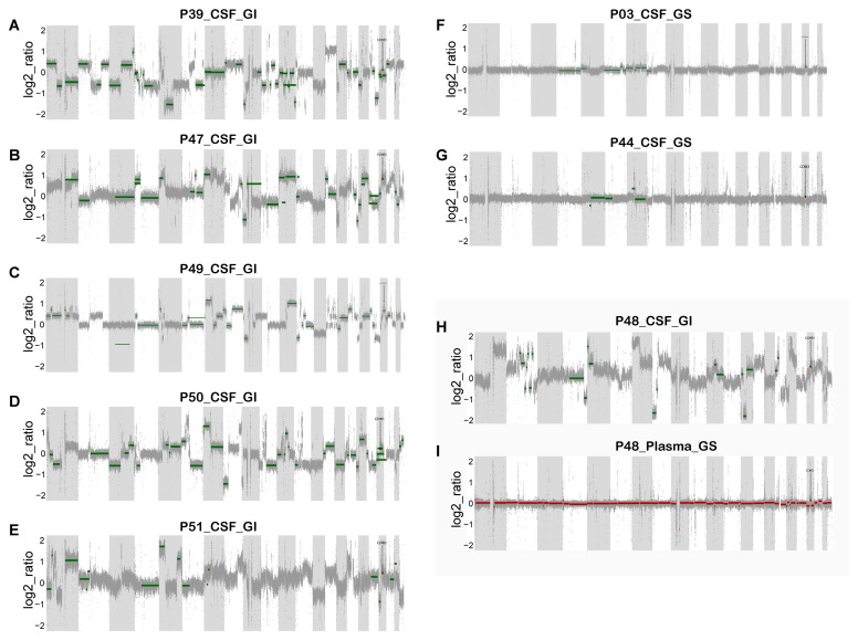
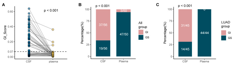
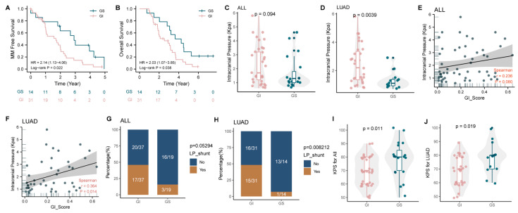
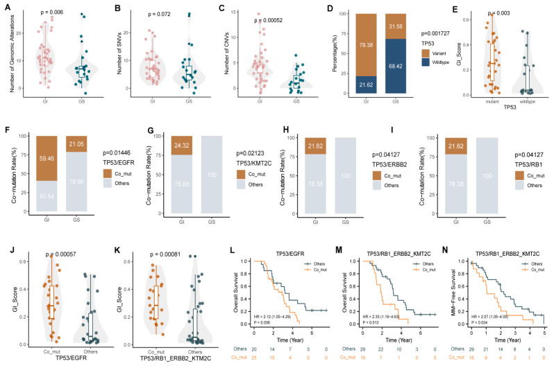
Genomic instability (GI), which leads to the accumulation of DNA loss, gain, and rearrangement, is a hallmark of many cancers such as lung cancer, breast cancer, and colon cancer. However, the clinical significance of GI has not been systematically studied in the meningeal metastasis (MM) of solid tumors. Here, we collected both cerebrospinal fluid (CSF) and plasma samples from 56 solid tumor MM patients and isolated cell-free ctDNA to investigate the GI status using a next-generation sequencing-based comprehensive genomic profiling of 543 cancer-related genes. According to the unfiltered heterozygous mutation data-derived GI score, we found that 37 (66.1%) cases of CSF and 3 cases (6%) of plasma had a high GI status, which was further validated by low-depth whole-genome sequencing analysis. It is demonstrated that a high GI status in CSF was associated with poor prognosis, high intracranial pressure, and low Karnofsky performance status scores. More notably, a high GI status was an independent poor prognostic factor of poor MM-free survival and overall survival in lung adenocarcinoma MM patients. Furthermore, high occurrences of the co-mutation of TP53/EGFR, TP53/RB1, TP53/ERBB2, and TP53/KMT2C were found in MM patients with a high GI status. In summary, the GI status in CSF ctDNA might be a valuable prognostic indicator in solid tumor patients with MM.

Keywords: cell-free DNA; cerebrospinal fluid; genomic instability; meningeal metastases; plasma.


Conflict of interest statement

The authors declare no conflict of interest.

Figures

Figure 1
Figure 1
CSF ctDNA is more reflective of the genetic profile of meningeal metastasis than plasma ctDNA. (A,B) Oncoprint of CSF (A) and plasma (B). Each column represents a patient; each row represents a gene. Different types of mutations are colored differently. The top bar represents the total number of mutations that a patient has. A sidebar summarizes the frequency of mutations in this cohort. (C,D) The total number of genetic variants (C) and the total number of SNVs (D) in CSF and plasma. (E,F) The detection rates of EGFR mutations (E) and TP53 mutations (F). (G) The detection rate is defined as having any copy number variation detected from the panel. (H) Copy numbers of genes exhibiting CNV in both CSF and plasma.
Figure 2
Figure 2
GS analysis of CSF and plasma based on a panel of 543 cancer-associated gene panels. The left side of the figure shows the allele frequency of shared mutations in leukocytes, plasma ctDNA and CSF ctDNA analyzed with unfiltered mutation data. The right side of the figure shows the distribution of the CSF gene copy number log2 ratio. All horizontal coordinates are chromosome locations. (AE) Results of five patients with GI. (FI) Results of four patients with GS. BC, white blood cells; CF, plasma; CSF, cerebrospinal fluid; CN, copy number.
Figure 3
Figure 3
The reliability of the GI score was successfully verified by low-depth WGS copy number analysis in eight patients. (AE,H) Changes in the gene copy number in the CSF of six patients with GI. (F,G) Changes in the gene copy number in the CSF of two patients with GS. (I) Changes in the gene copy number in the paired plasma of a genome-stable patient (P48).
Figure 4
Figure 4
MM is associated with increased GI. (A) GI scores in the GI and GS groups. (B,C) The proportions of 56 solid tumor MM patients (B) and 45 LUAD MM patients (C) with GI in CSF and plasma ctDNA.
Figure 5
Figure 5
GI is associated with a poor prognosis, a low Karnofsky functional status score, and severe intracranial hypertension. (A,B) Analysis of MFS (A) and OS (B) in genomically unstable and genomically stable LUAD patients with MM. (C,D) Intracranial pressure in the GI and GS groups for all patients and (D) for 45 patients with LUAD (C). (E,F) Correlation analysis between intracranial pressure and GI scores. (G,H) The proportion of patients who have had LP shunts in the GI and GS groups. (I,J) KPS scores in the GI and GS groups. MM, meningeal metastasis; LUAD, lung adenocarcinoma; KPS, Karnofsky performance status; LP, Lumboperitoneal.
Figure 6
Figure 6
Comparison of gene characteristics between the GI and GS groups and their relationship with GI and prognosis. (AC) The total number of gene mutations (A), the number of SNVs (B), and the number of CNVs (C) in the GI and GS groups. (D) Mutation rates of TP53 in the GI and GS groups. (E) GI scores in CSF samples with/without TP53 mutation. (FI) Detection of co-mutations of TP53/EGFR (F), TP53/KMT2C (G), TP53/ERBB2 (H), and TP53/RB1 (I) in the GI and GS groups. (J,K) GI scores in CSF samples with/without corresponding TP53 co-mutations. (LN) The presence of TP53 co-mutations was significantly associated with MM and poor prognosis.

Similar articles

Cited by

References

    1. Chamberlain M., Soffietti R., Raizer J., Rudà R., Brandsma D., Boogerd W., Taillibert S., Groves M.D., le Rhun E., Junck L., et al. Leptomeningeal metastasis: A Response Assessment in Neuro-Oncology critical review of endpoints and response criteria of published randomized clinical trials. Neuro-Oncology. 2014;16:1176–1185. doi: 10.1093/neuonc/nou089. - DOI - PMC - PubMed
    1. le Rhun E., Taillibert S., Chamberlain M.C. Carcinomatous meningitis: Leptomeningeal metastases in solid tumors. Surg. Neurol. Int. 2013;4:S265–S288. - PMC - PubMed
    1. Corbin Z.A., Nagpal S. Leptomeningeal Metastases. JAMA Oncol. 2016;2:839. doi: 10.1001/jamaoncol.2015.3502. - DOI - PubMed
    1. Yamaguchi H., Wakuda K., Fukuda M., Kenmotsu H., Mukae H., Ito K., Chibana K., Inoue K., Miura S., Tanaka K., et al. A Phase II Study of Osimertinib for Radiotherapy-Naive Central Nervous System Metastasis From NSCLC: Results for the T790M Cohort of the OCEAN Study (LOGIK1603/WJOG9116L) J. Thorac. Oncol. 2021;16:2121–2132. doi: 10.1016/j.jtho.2021.07.026. - DOI - PubMed
    1. Lee E., Keam B., Kim D.W., Kim T.M., Lee S.H., Chung D.H., Heo D.S. Erlotinib versus gefitinib for control of leptomeningeal carcinomatosis in non-small-cell lung cancer. J. Thorac. Oncol. 2013;8:1069–1074. doi: 10.1097/JTO.0b013e318294c8e8. - DOI - PubMed