

An Expanded Interplay Network between NF-κB p65 (RelA) and E2F1 Transcription Factors: Roles in Physiology and Pathology
- PMID: 36291831
- PMCID: PMC9600032
- DOI: 10.3390/cancers14205047
An Expanded Interplay Network between NF-κB p65 (RelA) and E2F1 Transcription Factors: Roles in Physiology and Pathology
Abstract
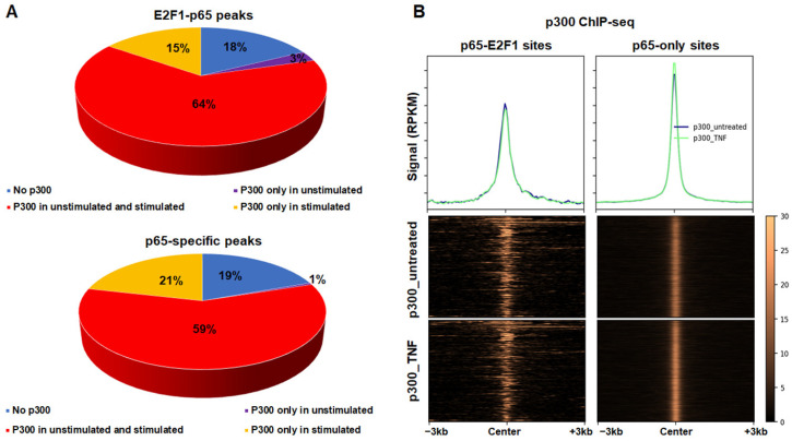
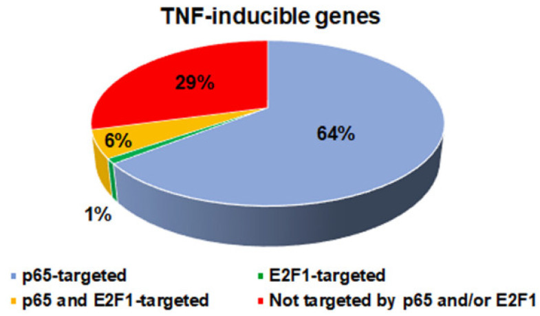
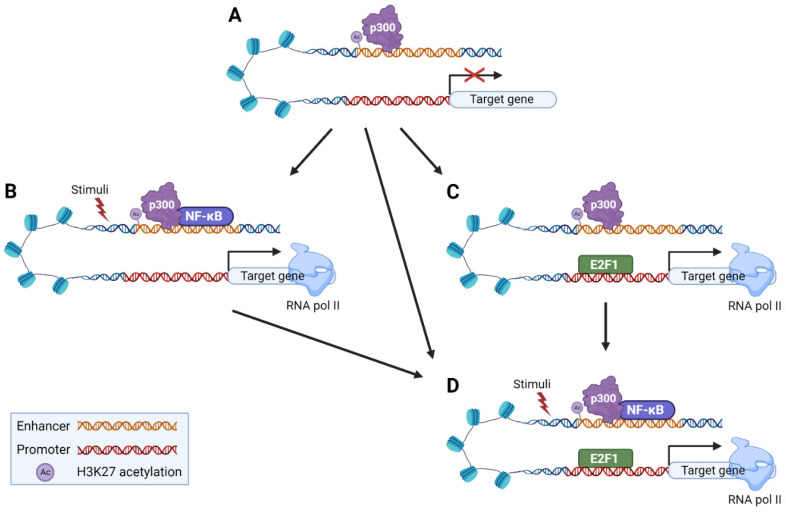
Transcription Factors (TFs) are the main regulators of gene expression, controlling among others cell homeostasis, identity, and fate. TFs may either act synergistically or antagonistically on nearby regulatory elements and their interplay may activate or repress gene expression. The family of NF-κB TFs is among the most important TFs in the regulation of inflammation, immunity, and stress-like responses, while they also control cell growth and survival, and are involved in inflammatory diseases and cancer. The family of E2F TFs are major regulators of cell cycle progression in most cell types. Several studies have suggested the interplay between these two TFs in the regulation of numerous genes controlling several biological processes. In the present study, we compared the genomic binding landscape of NF-κB RelA/p65 subunit and E2F1 TFs, based on high throughput ChIP-seq and RNA-seq data in different cell types. We confirmed that RelA/p65 has a binding profile with a high preference for distal enhancers bearing active chromatin marks which is distinct to that of E2F1, which mostly generates promoter-specific binding. Moreover, the RelA/p65 subunit and E2F1 cistromes have limited overlap and tend to bind chromatin that is in an active state even prior to immunogenic stimulation. Finally, we found that a fraction of the E2F1 cistrome is recruited by NF-κΒ near pro-inflammatory genes following LPS stimulation in immune cell types.
Keywords: E2F1; NF-κB; genomics; lung cancer; transcription factor interactions/interplay.
Conflict of interest statement
The authors declare no conflict of interest.
Figures


Similar articles
-
Genome-wide mapping of RELA(p65) binding identifies E2F1 as a transcriptional activator recruited by NF-kappaB upon TLR4 activation.Mol Cell. 2007 Aug 17;27(4):622-35. doi: 10.1016/j.molcel.2007.06.038. Mol Cell. 2007. PMID: 17707233
-
The interplay between NF-kappaB and E2F1 coordinately regulates inflammation and metabolism in human cardiac cells.PLoS One. 2011;6(5):e19724. doi: 10.1371/journal.pone.0019724. Epub 2011 May 23. PLoS One. 2011. PMID: 21625432 Free PMC article.
-
Transcriptional regulation of microRNA-100, -146a, and -150 genes by p53 and NFκB p65/RelA in mouse striatal STHdh(Q7)/ Hdh(Q7) cells and human cervical carcinoma HeLa cells.RNA Biol. 2015;12(4):457-77. doi: 10.1080/15476286.2015.1014288. RNA Biol. 2015. PMID: 25757558 Free PMC article.
-
New molecular bridge between RelA/p65 and NF-κB target genes via histone acetyltransferase TIP60 cofactor.J Biol Chem. 2012 Mar 2;287(10):7780-91. doi: 10.1074/jbc.M111.278465. Epub 2012 Jan 16. J Biol Chem. 2012. PMID: 22249179 Free PMC article.
-
Roles of NF-κB Signaling in the Regulation of miRNAs Impacting on Inflammation in Cancer.Biomedicines. 2018 Mar 30;6(2):40. doi: 10.3390/biomedicines6020040. Biomedicines. 2018. PMID: 29601548 Free PMC article. Review.
Cited by
-
Potential protective effects of Huanglian Jiedu Decoction against COVID-19-associated acute kidney injury: A network-based pharmacological and molecular docking study.Open Med (Wars). 2023 Jul 6;18(1):20230746. doi: 10.1515/med-2023-0746. eCollection 2023. Open Med (Wars). 2023. PMID: 37533739 Free PMC article.
References
Grants and funding
LinkOut - more resources
Full Text Sources
