. 2022 Sep 24;12(10):2303.
doi: 10.3390/diagnostics12102303.

Electric Source Imaging in Presurgical Evaluation of Epilepsy: An Inter-Analyser Agreement Study

Affiliations

Electric Source Imaging in Presurgical Evaluation of Epilepsy: An Inter-Analyser Agreement Study

Pietro Mattioli et al. Diagnostics (Basel). .

Abstract

Electric source imaging (ESI) estimates the cortical generator of the electroencephalography (EEG) signals recorded with scalp electrodes. ESI has gained increasing interest for the presurgical evaluation of patients with drug-resistant focal epilepsy. In spite of a standardised analysis pipeline, several aspects tailored to the individual patient involve subjective decisions of the expert performing the analysis, such as the selection of the analysed signals (interictal epileptiform discharges and seizures, identification of the onset epoch and time-point of the analysis). Our goal was to investigate the inter-analyser agreement of ESI in presurgical evaluations of epilepsy, using the same software and analysis pipeline. Six experts, of whom five had no previous experience in ESI, independently performed interictal and ictal ESI of 25 consecutive patients (17 temporal, 8 extratemporal) who underwent presurgical evaluation. The overall agreement among experts for the ESI methods was substantial (AC1 = 0.65; 95% CI: 0.59-0.71), and there was no significant difference between the methods. Our results suggest that using a standardised analysis pipeline, newly trained experts reach similar ESI solutions, calling for more standardisation in this emerging clinical application in neuroimaging.

Keywords: EEG; epilepsy; presurgical evaluation; source analysis; source imaging.


Conflict of interest statement

Dario Arnaldi: received fees from Fidia, Jazz and Lundbeck for lectures, consultation and board participation. All other authors declare no conflict of interest.

Figures
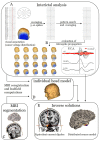
Figure 1
Figure 1
Analysis pipeline for interictal ESI. (A) First, a minimum of five IEDs of the same cluster (i.e., having the same voltage distribution) are visually annotated. Averaging of these IEDs creates a template which is used to perform a pattern search throughout the EEG file. The automatically detected IEDs are averaged to result in a better signal-to-noise ratio. (B) Next, intraspike propagation is assessed by visual analysis of the sequential voltage maps and a principal component analysis of the signal. (C) The patient’s MRI is segmented into different tissue classes; the electrodes are aligned to the scalp and are used to solve the forward problem, i.e., the creation of an individual head model (D). (E) Finally, source modelling is performed using two different inverse solution methods: equivalent current dipoles (ECD); a distributed source model (DSM), cortical CLARA.
Figure 2
Figure 2
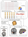
Analysis pipeline for ictal ESI. (A) The electrographic onset of the seizure is identified and marked manually. The first ictal waves with the same voltage distribution but without artefacts are annotated at their peaks. Next, these waves are averaged to increase the signal-to-noise ratio. (B) As these signals contain more noise than the interictal averaged signal, the signal is analysed at the peak. (C) The patient’s MRI is segmented into different tissue classes; the electrodes are aligned to the scalp and are used to solve the forward problem, i.e., the creation of an individual head model (D). (E) Finally, source modelling is performed using two different inverse solution methods: equivalent current dipoles (ECD); a distributed source model (DSM), cortical CLARA.
Figure 3
Figure 3
Examples of temporal (A) and extratemporal (B) sources. (A) Interictal LTM, ictal and interictal HD sources from a patient with temporal lobe epilepsy. The first row shows in a coronal plane the resulting dipoles of the ECD, which revealed a source in the right temporal pole. The second row depicts a cortical rendering of the patient’s MRI, seen in the right hemisphere. The result of the DSM also reveals a source in the right temporal pole. (B) Interictal LTM, ictal and interictal HD sources from a patient with extratemporal lobe epilepsy who previously had a resection in the right parietal lobe. The results of the ECD (first row, sagittal plane) and the DSM (second row, superior view on the cortical rendering) show sources around the previous resection cavity. (LTM: long-term monitoring, HD: high-density EEG, ECD: equivalent current dipole modelling, DSM: distributed source modelling).
Figure 4
Figure 4
The interval plot shows the agreement level of each ESI modality. On the y axis, AC values and 95% confidence intervals are reported. On the x axis, ESI modalities are reported. Analysers obtained substantial agreement in all ESIs modalities. Agreement level: <0: poor, 0.01–0.2: slight, 0.2–0.4: good, 0.4–0.6: moderate, 0.6–0.8: substantial, 0.8–1: almost perfect. The colour map underlines the level of agreement from red (poor) to dark green (almost perfect). (AC: agreement coefficient, ECD: equivalent current dipole, DSM: distributed source model, HD: high density, LTM: long term monitoring).

References

    1. Kwan P., Arzimanoglou A., Berg A.T., Brodie M.J., Allen Hauser W., Mathern G., Moshé S.L., Perucca E., Wiebe S., French J. Definition of drug resistant epilepsy: Consensus proposal by the ad hoc Task Force of the ILAE Commission on Therapeutic Strategies. Epilepsia. 2010;51:1069–1077. doi: 10.1111/j.1528-1167.2009.02397.x. - DOI - PubMed
    1. DeGiorgio C.M., Markovic D., Mazumder R., Moseley B.D. Ranking the Leading Risk Factors for Sudden Unexpected Death in Epilepsy. Front. Neurol. 2017;8:473. doi: 10.3389/fneur.2017.00473. - DOI - PMC - PubMed
    1. Ryvlin P., Cross H., Rheims S. Epilepsy surgery in children and adults. Lancet Neurol. 2014;13:1114–1126. doi: 10.1016/S1474-4422(14)70156-5. - DOI - PubMed
    1. Kwan P., Brodie M.J. Early Identification of Refractory Epilepsy. N. Engl. J. Med. 2000;342:314–319. doi: 10.1056/NEJM200002033420503. - DOI - PubMed
    1. Rosenow F., Lüders H. Presurgical evaluation of epilepsy. Brain. 2001;124:1683–1700. doi: 10.1093/brain/124.9.1683. - DOI - PubMed

LinkOut - more resources