

Case Reports
doi: 10.3390/diagnostics12102311.
The Onset of Subtalar Joint Monoarthritis in a Patient with Rheumatoid Arthritis
Affiliations
- PMID: 36292000
- PMCID: PMC9600197
- DOI: 10.3390/diagnostics12102311
Item in Clipboard
Case Reports
The Onset of Subtalar Joint Monoarthritis in a Patient with Rheumatoid Arthritis
Diagnostics (Basel).
.
Abstract
The involvement of the subtalar joint is uncommon in the early stages of rheumatoid arthritis (RA). We report a case of a 47-year-old female who had RA with isolated subtalar joint arthritis. The clinical history, magnetic resonance imaging, and pathological findings of the patient are presented. A careful evaluation of the patients for chronic ankle-to-heel pain should be conducted, and concomitant evaluation for inflammatory arthritis, including RA, should be considered.
Keywords: monoarthritis; rheumatoid arthritis; subtalar joint.
Conflict of interest statement
The authors declare no conflict of interest.
Figures

Subtalar joint involvement in a 47-year-old female. Anteroposterior (a) and lateral (b) plain radiographs of the right ankle joint. Coronal T1-weighted (c), sagittal T1-weighted (d), coronal fat-suppressed T2-weighted (e), and sagittal fat-suppressed T2-weighted (f) images of the right ankle joint using magnetic resonance imaging (MRI). MRI images show a moderate subtalar joint effusion with bone marrow edema.

Subtalar joint showing progressive involvement 1 year later. Coronal T1-weighted (a), sagittal T1-weighted (b), coronal fat-suppressed T2-weighted (c), and sagittal fat-suppressed T2-weighted (d) images of the right ankle joint. Findings show a marked loss of articular cartilage with associated bone marrow edema in the subarticular region of the subtalar joint and bone marrow edema of the tibiotalar joint.
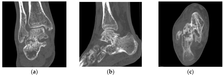
Erosion and destruction of the subtalar joint on computed tomography. Computed tomography images of the coronal (a), sagittal (b), and axial (c) parts of the subtalar joint. Findings demonstrate narrowed joint space and destruction of the subtalar joint of the right ankle.

Surgical biopsy specimen of the subtalar joint region shows synovial proliferation and lymphocytes in the synovium (a). Proliferation of synovial lining cells, vascular proliferation, and lymphocyte infiltrate can be observed in the synovium (b).
Similar articles
-
Do ankle, hindfoot, and heel ultrasound findings predict the symptomatology and quality of life in rheumatoid arthritis patients?J Ultrason. 2020;20(81):e70-e82. doi: 10.15557/JoU.2020.0012. Epub 2020 Jun 15. J Ultrason. 2020. PMID: 32609963 Free PMC article.
-
Relationship of ankle joint involvement with subtalar destruction in patients with rheumatoid arthritis. A 20-year follow-up study.Joint Bone Spine. 2001 Mar;68(2):154-7. doi: 10.1016/s1297-319x(00)00242-6. Joint Bone Spine. 2001. PMID: 11324931
-
Joint Distribution and Two-Year Outcome in 347 Patients With Monoarthritis of Less Than Sixteen Weeks' Duration.Arthritis Care Res (Hoboken). 2020 May;72(5):705-710. doi: 10.1002/acr.23334. Epub 2020 Apr 8. Arthritis Care Res (Hoboken). 2020. PMID: 28777897
-
The clinical features of rheumatoid arthritis.Eur J Radiol. 1998 May;27 Suppl 1:S18-24. doi: 10.1016/s0720-048x(98)00038-2. Eur J Radiol. 1998. PMID: 9652497 Review.
-
Natural history and imaging of subtalar and midfoot joint disease in rheumatoid arthritis.Int J Rheum Dis. 2013 Feb;16(1):14-8. doi: 10.1111/1756-185X.12035. Epub 2013 Feb 5. Int J Rheum Dis. 2013. PMID: 23441767 Review.
References
Publication types
LinkOut - more resources
Full Text Sources
