

Ultrasonographical Evaluation of the Median Nerve Mobility in Carpal Tunnel Syndrome: A Systematic Review and Meta-Analysis
- PMID: 36292039
- PMCID: PMC9600711
- DOI: 10.3390/diagnostics12102349
Ultrasonographical Evaluation of the Median Nerve Mobility in Carpal Tunnel Syndrome: A Systematic Review and Meta-Analysis
Abstract
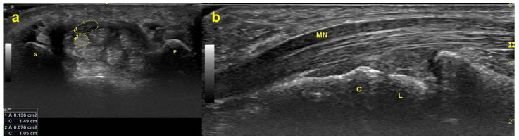
Diagnostic ultrasound is widely used for evaluating carpal tunnel syndrome (CTS), an entrapment neuropathy of the median nerve (MN). Decreased mobility of the MN inside the carpal tunnel has been reported in CTS, and various methods have been used to evaluate MN mobility; however, there is still no conclusive understanding of its connection with CTS. The purpose of this study is to conduct a systematic review and meta-analysis of the current published literature on ultrasonographic evaluations of transverse and longitudinal MN displacement and to identify the relationship between MN mobility and CTS. This study was conducted in accordance with the 2020 PRISMA statement and the Cochrane Collaboration Handbook. Comparative studies that investigated differences in MN displacement between CTS patients and healthy controls were retrieved by searching the Cochrane Library, Embase and PubMed. A total of 15 case-control studies were included. Nine of 12 studies evaluating transverse MN displacement and 4 of 5 studies evaluating longitudinal MN gliding showed that the MN was less mobile in CTS patients than in healthy subjects. Despite the large heterogeneity among the 15 included studies, this systematic review and meta-analysis provide evidence that the mobility of the MN is significantly reduced in both transverse and longitudinal planes in CTS patients compared to healthy controls. Five of the 15 included studies reported that a decrease in transverse or longitudinal MN displacement in CTS was correlated with clinical symptoms or with severity as measured by a nerve conduction study (NCS).
Keywords: carpal tunnel syndrome; median nerve; meta-analysis; nerve displacement; systematic review; ultrasound images.
Conflict of interest statement
The authors declare no conflict of interest.
Figures


Similar articles
-
Impaired median nerve mobility in patients with carpal tunnel syndrome: a systematic review and meta-analysis.Eur Radiol. 2023 Apr;33(4):2378-2385. doi: 10.1007/s00330-022-09262-9. Epub 2022 Nov 17. Eur Radiol. 2023. PMID: 36394604
-
Dexamethasone versus Hyaluronidase as an Adjuvant to Local Anesthetics in the Ultrasound-guided Hydrodissection of the Median Nerve for the Treatment of Carpal Tunnel Syndrome Patients.Anesth Essays Res. 2019 Jul-Sep;13(3):417-422. doi: 10.4103/aer.AER_104_19. Anesth Essays Res. 2019. PMID: 31602055 Free PMC article.
-
Ultrasonographic Measurement of the Median Nerve Transverse Diameter at the Wrist for Diagnosing Carpal Tunnel Syndrome.J Hand Surg Asian Pac Vol. 2021 Jun;26(2):223-228. doi: 10.1142/S2424835521500223. J Hand Surg Asian Pac Vol. 2021. PMID: 33928849
-
Quantitative Evaluation of the Echo Intensity of Paraneural Area and Myofascial Structure around Median Nerve in Carpal Tunnel Syndrome.Diagnostics (Basel). 2020 Nov 8;10(11):914. doi: 10.3390/diagnostics10110914. Diagnostics (Basel). 2020. PMID: 33171617 Free PMC article.
-
A Comparative Analysis Between Ultrasound and Electromyographic and Nerve Conduction Studies in Diagnosing Carpal Tunnel Syndrome (CTS): A Systematic Review and Meta-Analysis.Cureus. 2022 Oct 19;14(10):e30476. doi: 10.7759/cureus.30476. eCollection 2022 Oct. Cureus. 2022. PMID: 36415360 Free PMC article. Review.
Cited by
-
Successful evaluation of a new image-based parameter for the diagnosis of carpal tunnel syndrome: ultrasound assessment of longitudinal median nerve gliding in patients, healthy volunteers, and cadavers.Eur J Phys Rehabil Med. 2024 Aug;60(4):671-679. doi: 10.23736/S1973-9087.24.08491-0. Epub 2024 Jul 15. Eur J Phys Rehabil Med. 2024. PMID: 39007786 Free PMC article.
References
Grants and funding
LinkOut - more resources
Full Text Sources
Research Materials
