Comparison of Spectral-Domain OCT versus Swept-Source OCT for the Detection of Deep Optic Disc Drusen
- PMID: 36292204
- PMCID: PMC9600200
- DOI: 10.3390/diagnostics12102515
Comparison of Spectral-Domain OCT versus Swept-Source OCT for the Detection of Deep Optic Disc Drusen
Abstract
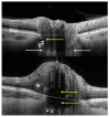
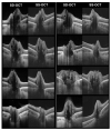
Deep optic disc drusen (ODD) are located below Bruch's membrane opening (BMO) and may go undetected due to the challenges in imaging them. The purpose of this study is a head-to-head comparison of currently widely used imaging technologies: swept-source optical coherence tomography (SS-OCT; DRI OCT-1 Triton, Topcon) and enhanced depth imaging spectral-domain optical coherence tomography (EDI SD-OCT; Spectralis OCT, Heidelberg Engineering) for the detection of deep ODD and associated imaging features. The eyes included in this study had undergone high-resolution imaging via both EDI SD-OCT and SS-OCT volume scans, which showed at least one deep ODD or a hyperreflective line (HL). Grading was performed by three graders in a masked fashion. The study findings are based on 46 B-scan stacks of 23 eyes including a total of 7981 scans. For scan images with ODD located above or below the level of BMO, no significant difference was found between the two modalities compared in this study. However, for HLs and other features, EDI SD-OCT scan images had better visualization and less artifacts. Although SS-OCT offers deep tissue visualization, it did not appear to offer any advantage in ODD detection over a dense volume scan via EDI SD-OCT with B-scan averaging.
Keywords: comparison; enhanced depth imaging; optic disc drusen; optic nerve head drusen; optical coherence tomography; spectral-domain; swept-source.
Conflict of interest statement
The authors declare no conflict of interest. The funders had no role in the design of the study; in the collection, analyses, or interpretation of data; in the writing of the manuscript; or in the decision to publish the results.
Figures


References
-
- Lorentzen S.E. Drusen of the Optic Disk. A Clinical and Genetic Study. Acta Ophthalmol. 1966;90:1–181. - PubMed
Grants and funding
LinkOut - more resources
Full Text Sources

