Augmented Reality Surgical Navigation System for External Ventricular Drain
- PMID: 36292263
- PMCID: PMC9601392
- DOI: 10.3390/healthcare10101815
Augmented Reality Surgical Navigation System for External Ventricular Drain
Abstract
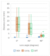
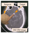
Augmented reality surgery systems are playing an increasing role in the operating room, but applying such systems to neurosurgery presents particular challenges. In addition to using augmented reality technology to display the position of the surgical target position in 3D in real time, the application must also display the scalpel entry point and scalpel orientation, with accurate superposition on the patient. To improve the intuitiveness, efficiency, and accuracy of extra-ventricular drain surgery, this paper proposes an augmented reality surgical navigation system which accurately superimposes the surgical target position, scalpel entry point, and scalpel direction on a patient's head and displays this data on a tablet. The accuracy of the optical measurement system (NDI Polaris Vicra) was first independently tested, and then complemented by the design of functions to help the surgeon quickly identify the surgical target position and determine the preferred entry point. A tablet PC was used to display the superimposed images of the surgical target, entry point, and scalpel on top of the patient, allowing for correct scalpel orientation. Digital imaging and communications in medicine (DICOM) results for the patient's computed tomography were used to create a phantom and its associated AR model. This model was then imported into the application, which was then executed on the tablet. In the preoperative phase, the technician first spent 5-7 min to superimpose the virtual image of the head and the scalpel. The surgeon then took 2 min to identify the intended target position and entry point position on the tablet, which then dynamically displayed the superimposed image of the head, target position, entry point position, and scalpel (including the scalpel tip and scalpel orientation). Multiple experiments were successfully conducted on the phantom, along with six practical trials of clinical neurosurgical EVD. In the 2D-plane-superposition model, the optical measurement system (NDI Polaris Vicra) provided highly accurate visualization (2.01 ± 1.12 mm). In hospital-based clinical trials, the average technician preparation time was 6 min, while the surgeon required an average of 3.5 min to set the target and entry-point positions and accurately overlay the orientation with an NDI surgical stick. In the preparation phase, the average time required for the DICOM-formatted image processing and program import was 120 ± 30 min. The accuracy of the designed augmented reality optical surgical navigation system met clinical requirements, and can provide a visual and intuitive guide for neurosurgeons. The surgeon can use the tablet application to obtain real-time DICOM-formatted images of the patient, change the position of the surgical entry point, and instantly obtain an updated surgical path and surgical angle. The proposed design can be used as the basis for various augmented reality brain surgery navigation systems in the future.
Keywords: augmented reality; neurosurgery; surgical navigation.
Conflict of interest statement
The authors declare no conflict of interest.
Figures


References
-
- Razzaque S., Heaney B. Medical Device Guidance. 10,314,559. US Patent. 2019 June 11;
-
- Bucholz D.R., Foley T.K., Smith K.R., Bass D., Wiedenmaier T., Pope T., Wiedenmaier U. Surgical Navigation Systems Including Reference and Localization Frames. 6,236,875. US Patent. 2001 May 22;
-
- Casas C.Q. Image-Guided Surgery with Surface Reconstruction and Augmented Reality Visualization. 10,154,239. US Patent. 2018 December 11;
Grants and funding
LinkOut - more resources
Full Text Sources
Research Materials
Miscellaneous

