

A Novel Transcriptome Approach to the Investigation of the Molecular Pathology of Vitreous and Retinal Detachment
- PMID: 36292771
- PMCID: PMC9601696
- DOI: 10.3390/genes13101885
A Novel Transcriptome Approach to the Investigation of the Molecular Pathology of Vitreous and Retinal Detachment
Abstract
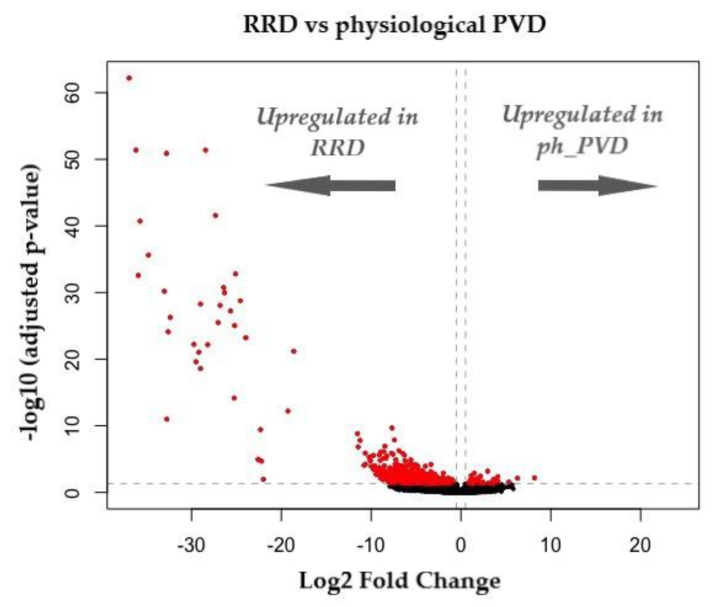
Retinal detachment (RD) is one of the most common, sight-threatening ocular conditions requiring emergency intervention. Posterior vitreous detachment (PVD) occurs in the majority of an aging population whereby the vitreous body separates from the retina. It is well established that PVD is the common precursor to the most common forms of RD; however, it remains unknown why in most individuals PVD will cause no/few complications (physiological PVD) but in a small percentage will cause retinal tears and detachment (pathological PVD). Despite over 100 years of scientific research, the anatomical definitions of PVD and its pathogenesis remain controversial. Recent research has identified a novel cell population (laminocyte), present at significantly higher numbers in pathological PVD when compared to physiological PVD. We review and summarise the seven distinct clinical sub-groups of retinal breaks and focus on the role of the laminocyte in those secondary to PVD and the transcriptomic profile of this unique cell. Provisional whole transcriptome analysis using bulk RNA-Seq shows marked differentially expressed genes when comparing physiological PVD with PVD associated with RD. The limitations of bulk RNA-Seq are considered and the potential to address these using spatial transcriptomics are discussed. Understanding the pathogenesis of PVD-related retinal tears will provide a baseline for the development of novel therapeutic targets and prophylactic treatments.
Keywords: RNA sequencing (RNA-seq); laminocyte; posterior hyaloid membrane (PHM); posterior vitreous detachment (PVD); retinal detachment (RD); rhegmatogenous retinal detachment (RRD); spatial transcriptomics.
Conflict of interest statement
The authors declare no conflict of interest.
Figures
References
-
- NHS Digital. Latest Figures on Registered Blind and Partially Sighted People in England are Published. 2021. [(accessed on 17 May 2022)]. Available online: https://digital.nhs.uk/news/2021/latest-figures-on-registered-blind-and-....
Publication types
MeSH terms
LinkOut - more resources
Full Text Sources
Medical
Miscellaneous
