Review
. 2022 Oct 21;13(10):1917.
doi: 10.3390/genes13101917.

Advances in Novel Animal Vitamin C Biosynthesis Pathways and the Role of Prokaryote-Based Inferences to Understand Their Origin

Affiliations
Review

Advances in Novel Animal Vitamin C Biosynthesis Pathways and the Role of Prokaryote-Based Inferences to Understand Their Origin

Pedro Duque et al. Genes (Basel). .

Abstract

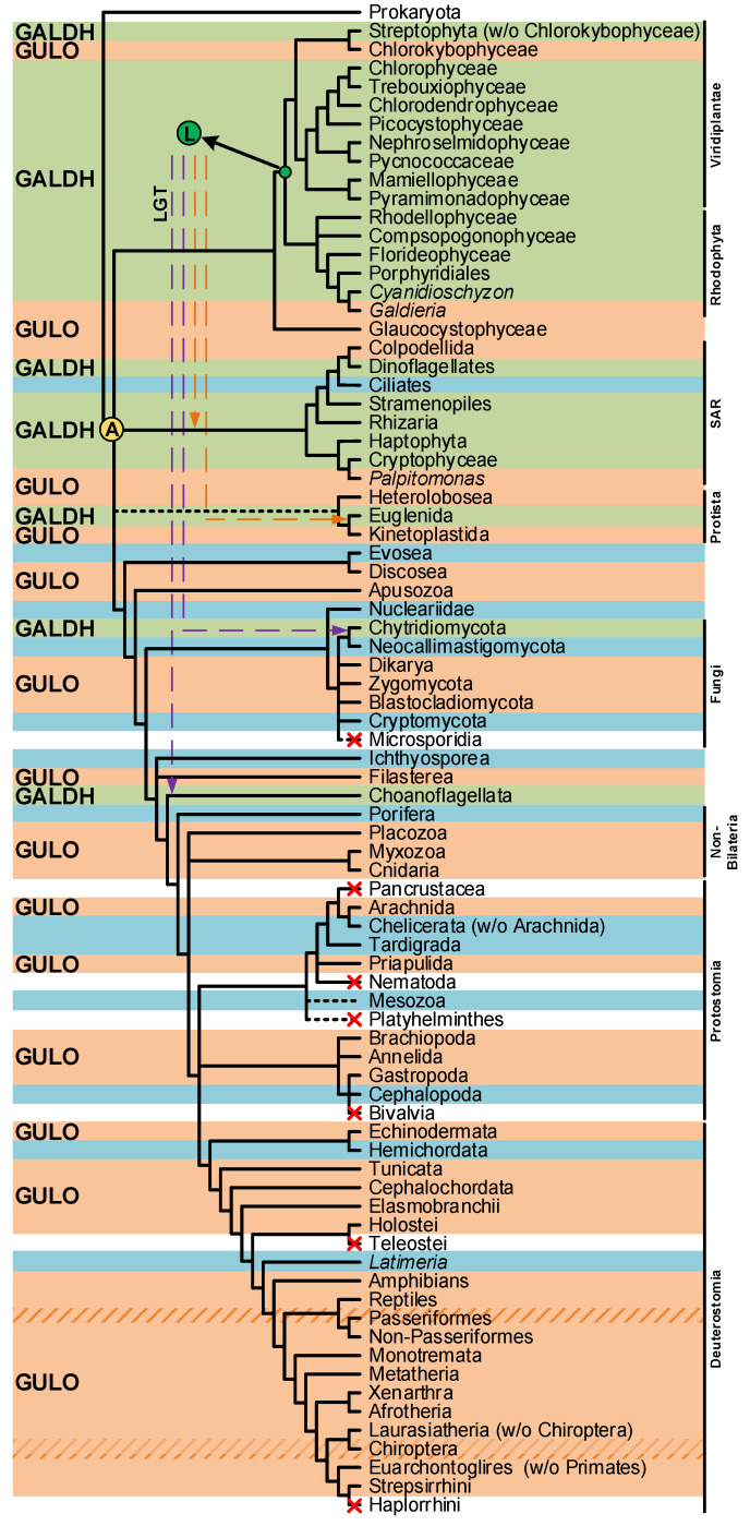
Vitamin C (VC) is an essential nutrient required for the optimal function and development of many organisms. VC has been studied for many decades, and still today, the characterization of its functions is a dynamic scientific field, mainly because of its commercial and therapeutic applications. In this review, we discuss, in a comparative way, the increasing evidence for alternative VC synthesis pathways in insects and nematodes, and the potential of myo-inositol as a possible substrate for this metabolic process in metazoans. Methodological approaches that may be useful for the future characterization of the VC synthesis pathways of Caenorhabditis elegans and Drosophila melanogaster are here discussed. We also summarize the current distribution of the eukaryote aldonolactone oxidoreductases gene lineages, while highlighting the added value of studies on prokaryote species that are likely able to synthesize VC for both the characterization of novel VC synthesis pathways and inferences on the complex evolutionary history of such pathways. Such work may help improve the industrial production of VC.

Keywords: aldonolactone oxidoreductases; ascorbic acid; evolution; insects; nematodes; prokaryotes; synthesis.


Conflict of interest statement

The authors declare no conflict of interest.

Figures

Figure 1
Figure 1
Graphical illustration of the main VC and D-erythroascorbate synthesis pathways, adapted from the MetaCyc database [108]. The numbered circles represent enzymes that are known to catalyze the substrate conversions within the pathways (see the enzyme list presented below the illustration). Reaction steps without an attributed enzyme are performed by unknown enzymes.
Figure 2
Figure 2
Inferences on enzymatic mechanisms and possible substrates utilized by C. elegans and D. melanogaster in the VC animal synthesis pathways. The ellipses highlight the main substrates possibly used in the proposed pathways, but that remain to be addressed using appropriate experimental setups. Enzymes that may be correlated with VC synthesis in these species are highlighted in bold next to the metabolic steps they are likely to catalyze. The interrogation marks indicate uncertainty in the steps regarding mechanisms of conversion of uncharacterized substrates, and the relative contribution of these processes to the availability of VC precursors. Common sugars (such as D-mannose, L-Galactose, D-Glucose and D-Arabinose) are assigned with a while L-Galactono-1,4-lactone, L-Gulono-1,4-lactone and D-Arabinono-1,4-lactone are assigned with b.
Figure 3
Figure 3
Presence/absence pattern of the GULO/GALDH lineages in eukaryotes. The taxonomic groups where the GULO lineage is present are highlighted by an orange background, while those where the GALDH lineage is present are highlighted by a green background. Independent GULO loss events within specific vertebrate groups are represented by stripe patterns. The blue background highlights uncertainty regarding either gene lineage presence/absence. These cases represent groups where both gene lineages could not be found in less than three species, or groups where the sequences identified were likely derived from genome contamination. Taxonomic groups where either gene lineage was lost are not highlighted, and a red cross can be seen in overlapping the branches they represent. Higher taxonomic ranks were added to the right of the figure to facilitate the interpretation. The ancestral paralogy and lateral gene transfer hypotheses proposed in [94] are indicated by the yellow and green filled circles with an A and L acronyms over the relevant tree nodes, respectively. The implied duplication events occurred during the time scale represented by the branches that led to the highlighted nodes. Both gene lineages already existed in scenario A, and the pattern of loss/presence of each was based upon selective constraints of the distinct taxonomic groups. Scenario L implies the origin of the GALDH lineage in the common ancestor of the Rhodophyta and Viridiplantae, that was later transferred to other taxonomic groups by endosymbiotic (orange dashed arrows) or horizontal (dashed purple arrows) gene transfer events. The data depicted in the figure was obtained from [94,112,114], and the taxonomic relationships are based on those found in [194,195] and the Tree of Life Web Project (http://tolweb.org/tree/; [196]; last accessed on 12 October 2022).
Figure 4
Figure 4
Putative scenarios of eukaryote AO evolution derived from prokaryote LGT events. In panel (A), a prokaryote GUDH was initially integrated in the genome of the ancestral eukaryote after an EGT event. Two hypotheses for the divergence of GUDH in eukaryotes are presented. In the left (yellow background), the GUDH present in the ancestral eukaryote was duplicated, originating GUDH1 and GUDH2. Distinct selective pressures led to the loss of the GALDH ancestral gene (GUDH2) in glaucophytes and opisthokonts, and thus GULO is the result of GUDH1 evolution. In the non-glaucophyta Archaeplastida, GALDH was the result of selective pressures imposed on GUDH2, while GUDH1 maintained the ancestral state. During evolution, GUDH1 was eventually lost in taxonomic groups that diverged from the one where A. thaliana is included. This scenario is compatible with the ancient paralogy hypothesis presented in [94], as indicated in the figure. In the right (purple background), the scenario represents the direct origin of the opisthokont and glaucophyte GULO lineages as the result of selective pressures imposed on the ancestral GUDH. In this case, a GUDH duplication event affected only the Archaeplastida ancestor after the divergence from the glaucophytes, and the GUDH1 and GALDH duplicates are the result of distinct selective pressures. Then, GUDH1 was lost in taxonomic groups that diverged from the one where A. thaliana is included. This scenario is more compatible with the LGT hypothesis presented in [94], as indicated in the figure. In panel (B), GULO and ALO, in animals and fungi, respectively, are the result of selective pressures imposed on an AO acquired through HGT by the ancestral opisthokont.
Figure 5
Figure 5
Bayesian phylogenetic analysis of eukaryote and putative prokaryote AOs. Two DHCR24 sequences from Homo sapiens and B. mori were used to root the tree. The remaining sequences are colored according to the relevant taxonomic groups analyzed, following the legend available below the phylogeny. The likely type of AO represented in each inferred sequence cluster can be seen to the right of the phylogeny. The GUDH, GULO, GAL, GALDH and ALO acronyms are indicative of L-Gulono-1,4-lactone Dehydrogenase, L-Gulonolactone oxidase, L-Galactono-1,4-lactone oxidase, L-Galactono-1,4-lactone dehydrogenase and D-Arabinono-1,4-lactone oxidase, respectively. The bacterial and plant GUDHs are distinguished by the “b” and “p” prefixes, respectively. The sequences used for the phylogenetic inference were obtained using the “blastp” algorithm integrated in the NCBI database (https://blast.ncbi.nlm.nih.gov/Blast.cgi; accessed on 26 July 2022) and the S. cerevisiae S288C ALO sequence (NP_013624.1) as query against the genomes of Actinobacteria, Proteobacteria, Bacteroidetes, Cyanobacteria, Archaea, Viridiplantae, fungi and Euglenozoa species. This genome selection considered bacterial groups previously correlated with VC synthesis [87,88,89,90,91,92,93], as well as the complementation of prokaryote taxonomy by the inclusion of Archaea. The Viridiplantae, fungi and Euglenozoa groups were selected to provide methodological validation relative to current literature and overall phylogenetic context. Homologous sequences representative of two species of each bacterial group and Archaea, five species of Viridiplantae, five species of fungi and three species of Euglenozoa were extracted. The “blastp” output produced considerably more results for each taxonomic group than those extracted, but the restrictive selection is representative, and through the use of more elaborated methods that have limitations in relation to the number of sequences they can handle, the quality of the phylogenetic inferences was improved. In addition to these sequences, five animal GULO sequences identified in [112] were also included to facilitate the interpretation of the phylogenetic results, as well as two sterol reductases sequences encoding the human and B. mori DHCR24 proteins, to be used as outgroup. The SEDA software [219] was used to verify the datasets and remove any anomalous sequence or isoforms that could be present. The Bayesian phylogenetic inference was performed using a MUSCLE [220] alignment as implemented in T-Coffee [221] and 2,000,000 tree generations with a defined burn-in of 25% as parameters for MrBayes [222], within ADOPS [223]. For both SEDA and ADOPS, the Docker images that are available at the pegi3S Bioinformatics Docker Images Project (https://pegi3s.github.io/dockerfiles/; accessed on 26 July 2022; [224]) were used.
Figure 6
Figure 6
Summary of the findings discussed in this review. The taxonomic relationship between the species mentioned in this work is displayed on the top of the figure. The characterized VC synthesis pathways and the putative alternatives in eukaryote and prokaryote species are presented in the mid part of the figure. The pathways already described in the literature are highlighted with bold colors according to the taxonomic groups mentioned, while the putative VC pathways are presented in the gridline sections. The enzymes that intervene in the substrate conversions are presented by numbered circles according to the following scheme: Circles in white represent enzymatic reactions described in the literature, circles in yellow represent hypothetical enzymatic reactions and circles in green indicate novel enzymatic constituents of putative alternative VC synthesis pathways with available functional characterization. The question marks (?) represent putative enzymatic reactions for which no candidate enzyme can be suggested with current data. The tissues correlated with VC synthesis in animal species are represented below the corresponding proposed pathways when available. The dashed box highlights the myo-Inositol VC synthesis pathway that is exclusively used by species that synthesize VC in the kidney. The phylogenetic relationship inferred between the studied AOs is summarized in a cladogram format at the bottom. The enzyme number and substrate acronym lists are presented below the figure to facilitate the interpretation.

Similar articles

Cited by

References

    1. Du J., Cullen J.J., Buettner G.R. Ascorbic acid: Chemistry, biology and the treatment of cancer. Biochim. Biophys. Acta. 2012;1826:443–457. doi: 10.1016/j.bbcan.2012.06.003. - DOI - PMC - PubMed
    1. Bauernfeind J.C. Ascorbic Acid: Chemistry, Metabolism, and Uses. American Chemical Society; Washington, DC, USA: 1982. Ascorbic Acid Technology in Agricultural, Pharmaceutical, Food, and Industrial Applications; pp. 395–497. Advances in Chemistry.
    1. Yin X., Chen K., Cheng H., Chen X., Feng S., Song Y., Liang L. Chemical Stability of Ascorbic Acid Integrated into Commercial Products: A Review on Bioactivity and Delivery Technology. Antioxidants. 2022;11:153. doi: 10.3390/antiox11010153. - DOI - PMC - PubMed
    1. Chaturvedi S., Khan S., Bhunia R.K., Kaur K., Tiwari S. Metabolic engineering in food crops to enhance ascorbic acid production: Crop biofortification perspectives for human health. Physiol. Mol. Biol. Plants. 2022;28:871–884. doi: 10.1007/s12298-022-01172-w. - DOI - PMC - PubMed
    1. Hieu T.V., Guntoro B., Qui N.H., Quyen N.T.K., Hafiz F.A.A. The application of ascorbic acid as a therapeutic feed additive to boost immunity and antioxidant activity of poultry in heat stress environment. Vet. World. 2022;15:685–693. doi: 10.14202/vetworld.2022.685-693. - DOI - PMC - PubMed

Publication types

LinkOut - more resources