Meta-Analysis
. 2022 Oct 11;23(20):12113.
doi: 10.3390/ijms232012113.

Role of Ion Channels in the Maintenance of Sperm Motility and Swimming Behavior in a Marine Teleost

Affiliations
Meta-Analysis

Role of Ion Channels in the Maintenance of Sperm Motility and Swimming Behavior in a Marine Teleost

Júlia Castro-Arnau et al. Int J Mol Sci. .

Abstract

In oviparous marine fishes, the hyperosmotic induction of sperm motility in seawater (SW) is well established, however, the potential function of ion channels in the maintenance of post activated spermatozoon swimming performance remains largely unknown. Here, we investigated the influence of ion channels on the spermatozoon swimming parameters using the gilthead seabream (Sparus aurata) as a model for modern marine teleosts. Our data show that the SW-induced activation of seabream sperm motility requires three concomitant processes, the hyperosmotic shock, an ion-flux independent increase of the intracellular concentration of Ca2+ ([Ca2+]i), but not of [K+]i or [Na+]i, and the alkalization of the cytosol. The combination of all three processes is obligatory to trigger flagellar beating. However, the time-course monitoring of sperm motion kinetics and changes in the [Ca2+]i, [K+]i and [Na+]i in SW or in non-ionic activation media, showed that the post activated maintenance of spermatozoa motility is dependent on extracellular Ca2+ and K+. A meta-analysis of a seabream sperm transcriptome uncovered the expression of multiple ion channels, some of which were immunolocalized in the head and/or tail of the spermatozoon. Selective pharmacological inhibition of these ion channel families impaired the long-term motility, progressivity, and velocity of SW-activated spermatozoa. The data further revealed that some antagonists of K+-selective or Ca2+-selective channels, as well as of stretch-activated and mechanosensitive channels, altered the trajectory of spermatozoa, suggesting that these ion channels are likely involved in the control of the swimming pattern of the post activated spermatozoon. These combined findings provide new insight into the signaling pathways regulating spermatozoon activation and swimming performance in marine fishes.

Keywords: activation; ion channels; ions; motility; pH; spermatozoa; trajectory; transcriptome.


Conflict of interest statement

The authors declare no conflict of interest. The funders had no role in the design of the study; in the collection, analyses, or interpretation of data; in the writing of the manuscript, or in the decision to publish the results.

Figures

Figure 1
Figure 1
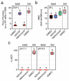
A surge of [Ca2+]i, but not [K+]i or [Na+]i, independent of external ions, is necessary for the activation of sperm motility in the seabream. (a) Effect of different incubations times in standard non-activating medium (NAM) or NAM containing sucrose and no ions (NAMsuc), and further activation in seawater (SW) or sucrose (Suc) for 5 s, on spermatozoa motility (% MOT), progressivity (% PROG), and curvilinear velocity (VCL). (b) Levels of [Ca2+]i, [K+]i and [Na+] at 5 s post activation in spermatozoa treated as in (a). (c) Percentage of MOT, and intracellular levels of Ca2+ and K+, in sperm activated in SW containing increasing doses of BAPTA. In all panels, the data points are presented as box and whisker plots/scatter dots with horizontal line (inside box) indicating median and outliers. One ejaculate from each male was measured from n = 4–7 males. Data were statistically analyzed by one-way ANOVA. Boxes with different superscript are statistically significant (p < 0.05). n.s., not significant.
Figure 2
Figure 2
A basic pH is necessary to activate and maintain motility of seabream spermatozoa. (a) Time-course of total motile sperm (% MOT) and curvilinear velocity (VCL) upon activation in seawater (SW) at pH 6 or 8. Sperm activated at pH 6 was exposed to 250 mM NH4Cl at 25 s after activation (arrowhead). (b) % MOT and VCL of spermatozoa activated in SW at constant pH 8, or exposed to 1.6 mM HCl at 25 s after activation (arrowheads). Data are the mean ± SEM (n = 3–4 males, one ejaculate per male).
Figure 3
Figure 3
High levels of intracellular Ca2+ and basic pH are not sufficient for the activation of motility of seabream spermatozoa. (a,b) Intracellular Ca2+ (a) and K+ (b) levels in immotile sperm treated with 25 mM of the Ca2+ ionophore A23187 or the K+ ionophore valinomycin for up to 30 min, and in SW-activated spermatozoa. (c) Percentage of motility (% MOT) of the sperm treated as above under neutral and basic pH. In all panels, the data points are presented as box and whisker plots/scatter dots with horizontal line (inside box) indicating median and outliers. One ejaculate from each male was measured from n = 4–6 males. Statistical differences were measured by one-way ANOVA. Boxes with different superscript are statistically significant (p < 0.05).
Figure 4
Figure 4
External ions are required for the maintenance of motility of post activated seabream spermatozoa. (a) Motility, progressivity, and VCL, of spermatozoa upon activation in SW or sucrose. (b) Intracellular Ca2+, K+ and Na+ levels in sperm activated in SW or sucrose. (c) Motility, progressivity, and VCL, of spermatozoa upon activation in SW or 550 mM NaCl. (d) Effect of activation in SW, NaCl, or NaCl, in which 10 mM Ca2+ and K+ were added separately or together, on seabream sperm kinetics. In a-c, data are the mean ± SEM (n = 4–5 males, one ejaculate per male), and were statistically analyzed by two-way ANOVA (p values indicated in each panel). In (d), data points (n = 4) are presented as box and whisker plots/scatter dots with horizontal line (inside box) indicating median and outliers. One ejaculate from each male was measured. Statistical differences were measured by one-way ANOVA for each post activation time point, followed by the Tukey’s multiple comparison test. Boxes with different superscript are statistically significant (p < 0.05). n.s., not significant.
Figure 5
Figure 5
Ion channel-encoding genes expressed in ejaculated seabream spermatozoa. (a) Plot showing the mean expression levels of all the 342 genes assessed as fragments per kilo base per million mapped reads (FPKM) of the corresponding transcripts. (b) Levels of expression of the most abundant transcripts encoding for ion channels potentially localized in the plasma membrane of the spermatozoa determined in 5 replicate RNA-seq libraries of ejaculated spermatozoa (each replicate being a pool of cells collected from three different males). Data points are presented as box and whisker plots/scatter dots with horizontal line (inside box) indicating median and outliers. Data in a and b were calculated from a recent RNA-seq analysis on seabream spermatozoa reported by Castro-Arnau et al. [30] (Gene Expression Omnibus database accession no. GSE173088, National Center for Biotechnology Information). (c) Representative RT-PCR detection of mRNAs encoding selected ion channels in testis (T) and ejaculated spermatozoa (S). The n line is the negative control (absence of RT during cDNA synthesis). The arrows indicate the specific amplified transcripts, and the size (kb) of molecular markers are indicated on the left. Uncropped gels are shown in Supplementary Figure S2). See Spreadsheets S1 and S2 for abbreviations and gene names.
Figure 6
Figure 6
Confirmation of protein expression of different ion channels in seabream immotile and activated spermatozoa. (ah, upper panels) Immunoblots of selected ion channels in immotile and motile spermatozoa (Si and Sm, respectively). Alpha-tubulin (Tuba) was used as a marker for even loading. Arrows indicate channel monomers and arrowheads the expected size of the target bands based on in silico determination of molecular masses. Asterisks indicate potential post translational modifications. Molecular mass markers (kDa) are on the left. Uncropped immunoblots are shown in Supplementary Figure S3. (ah, lower panels) Representative bright field (BF) images (left) and immunodetection (right; red color) of ion channels in ejaculated immotile spermatozoa. The spermatozoon nucleus is counterstained with DAPI (blue). Scale bars, 5 µm.
Figure 7
Figure 7
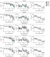
Inhibition of seabream sperm motility by blockers of Ca2+ channels, ATP-sensitive and voltage-dependent K+ channels, and CNG channels. (ao) Dose-response inhibition of the percentage of motility and progressivity (% MOT and % PROG, respectively) and curvilinear velocity (VCL) at 5, 30 and 60 s post activation induced by the different ion channel blockers as indicated. Control spermatozoa were treated with 0.5% DMSO. In all panels, the data points (n = 6–8 males, one ejaculated per male) are presented as box and whisker plots/scatter dots with horizontal line (inside box) indicating median and outliers. Statistical differences were measured by one-way ANOVA (*, p < 0.05; **, p < 0.01; ***, p < 0.001, with respect to DMSO-treated sperm).
Figure 8
Figure 8
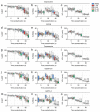
Inhibition of seabream sperm motility by blockers of voltage-gated Na+ channels, VRAC, TRPV, SACs, and MSCs. (ao) Dose-response inhibition of the percentage of motility and progressivity (% MOT and % PROG, respectively) and curvilinear velocity (VCL) at 5, 30 and 60 s post activation induced by the different ion channel blockers as indicated. Control spermatozoa were treated with 0.5% DMSO. In all panels, the data points (n = 6–8 males, one ejaculated per male) are presented as box and whisker plots/scatter dots with horizontal line (inside box) indicating median and outliers. Statistical differences were measured by one-way ANOVA (*, p < 0.05; **, p < 0.01; ***, p < 0.001, with respect to DMSO-treated sperm).
Figure 9
Figure 9
Percentage of inhibition of seabream sperm motility (MOT), progressivity (PROG), and curvilinear velocity (VCL), by the different ion channel blockers tested. (aj) The percentage of inhibition at each dose and post activation time (30 or 60 s) with respect to DMSO-treated spermatozoa was calculated from data shown in Figure 7 and Figure 8. In all panels, the data points (n = 6–8 males, one ejaculated per male) are presented as box and whisker plots/scatter dots with horizontal line (inside box) indicating median and outliers. The asterisks indicate statistical differences between the percentage of inhibition of PROG and MOT at the same dose and post activation time (Student t-test; *, p < 0.05; **, p < 0.01; ***, p < 0.001).
Figure 10
Figure 10
Effect of selected ion channel blockers on the trajectory of seabream spermatozoa. (af) Absolute mean angular displacement (MAD) of sperm treated with DMSO or the inhibitors at 30 or 60 s post activation in SW. Data points (n = 6–8 males, one ejaculated per male) are presented as box and whisker plots/scatter dots with horizontal line (inside box) indicating median and outliers. Statistical differences were measured by one-way ANOVA (*, p < 0.05; **, p < 0.01; ***, p < 0.001, with respect to DMSO-treated sperm). (gl) Representative trajectory of a single activated spermatozoon in the presence of DMSO or the different inhibitors analyzed with ISAS®v1 CASA-Mot system. The red line shows curvilinear velocity (VCL), the blue line shows straight line velocity (VSL), and the green line shows average path velocity (VAP). The complete capture video sequences tracked at 25 frames per second are shown in Supplementary Figure S1.

References

    1. Pitnick S., Wolfner M.F., Dorus S. Post-ejaculatory modifications to sperm (PEMS) Biol. Rev. Camb. Philos. Soc. 2020;95:365–392. doi: 10.1111/brv.12569. - DOI - PMC - PubMed
    1. Alavi S.M.H., Cosson J., Bondarenko O., Linhart O. Sperm motility in fishes: (III) diversity of regulatory signals from membrane to the axoneme. Theriogenology. 2019;136:143–165. doi: 10.1016/j.theriogenology.2019.06.038. - DOI - PubMed
    1. Cosson J., Groison A., Suquet M., Fauvel C., Dreanno C., Billard R. Studying sperm motility in marine fish: An overview on the state of the art. J. Appl. Ichthyol. 2008;24:460–486. doi: 10.1111/j.1439-0426.2008.01151.x. - DOI
    1. Darszon A., Nishigaki T., Beltran C., Treviño C.L. Calcium channels in the development, maturation, and function of spermatozoa. Physiol. Rev. 2011;91:1305–1355. doi: 10.1152/physrev.00028.2010. - DOI - PubMed
    1. Fechner S., Alvarez L., Bönigk W., Müller A., Berger T.K., Pascal R., Trötschel C., Poetsch A., Stölting G., Siegfried K.R., et al. A K+-selective CNG channel orchestrates Ca2+ signalling in zebrafish sperm. Elife. 2015;4:e07624. doi: 10.7554/eLife.07624. - DOI - PMC - PubMed

Publication types