Iron Deficiency Promotes the Lack of Photosynthetic Cytochrome c550 and Affects the Binding of the Luminal Extrinsic Subunits to Photosystem II in the Diatom Phaeodactylum tricornutum
- PMID: 36292994
- PMCID: PMC9603157
- DOI: 10.3390/ijms232012138
Iron Deficiency Promotes the Lack of Photosynthetic Cytochrome c550 and Affects the Binding of the Luminal Extrinsic Subunits to Photosystem II in the Diatom Phaeodactylum tricornutum
Abstract
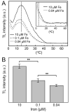
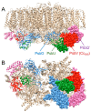
In the diatom Phaeodactylum tricornutum, iron limitation promotes a decrease in the content of photosystem II, as determined by measurements of oxygen-evolving activity, thermoluminescence, chlorophyll fluorescence analyses and protein quantification methods. Thermoluminescence experiments also indicate that iron limitation induces subtle changes in the energetics of the recombination reaction between reduced QB and the S2/S3 states of the water-splitting machinery. However, electron transfer from QA to QB, involving non-heme iron, seems not to be significantly inhibited. Moreover, iron deficiency promotes a severe decrease in the content of the extrinsic PsbV/cytochrome c550 subunit of photosystem II, which appears in eukaryotic algae from the red photosynthetic lineage (including diatoms) but is absent in green algae and plants. The decline in the content of cytochrome c550 under iron-limiting conditions is accompanied by a decrease in the binding of this protein to photosystem II, and also of the extrinsic PsbO subunit. We propose that the lack of cytochrome c550, induced by iron deficiency, specifically affects the binding of other extrinsic subunits of photosystem II, as previously described in cyanobacterial PsbV mutants.
Keywords: Phaeodactylum tricornutum; PsbV; cytochrome c550; electron transfer; iron limitation; photosystem II; pulse-amplitude modulation chlorophyll fluorescence; thermoluminescence.
Conflict of interest statement
The authors declare no conflict of interest.
Figures


References
-
- Kranzler C., Rudolf M., Keren N., Schleif E. Iron in Cyanobacteria. In: Chauvat F., Cassier-Chauvat C., editors. Genomics of Cyanobacteria. Volume 65. Academic Press; San Diego, CA, USA: 2013. pp. 57–105. Advances in Botanical Research.
-
- Geider R.J., La Roche J., Greene R.M., Olaizola M. Response of the photosynthetic apparatus of Phaeodactylum tricornutum (Bacillariophyceae) to nitrate, phosphate, or iron starvation. J. Phycol. 1993;29:755–766. doi: 10.1111/j.0022-3646.1993.00755.x. - DOI
-
- Moore J.K., Doney S.C., Glover D.M., Fung I.Y. Iron cycling and nutrient limitation patterns in surface waters of the world ocean. Deep Sea Res. Part II. 2002;49:463–508. doi: 10.1016/S0967-0645(01)00109-6. - DOI
MeSH terms
Substances
Grants and funding
LinkOut - more resources
Full Text Sources

