. 2022 Oct 14;23(20):12318.
doi: 10.3390/ijms232012318.

Binding of Glycerol to Human Galectin-7 Expands Stability and Modulates Its Functions

Affiliations

Binding of Glycerol to Human Galectin-7 Expands Stability and Modulates Its Functions

Yebing Liang et al. Int J Mol Sci. .

Abstract

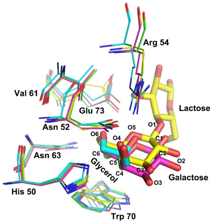
Glycerol is seen in biological systems as an intermediate in lipid metabolism. In recent years, glycerol has been reported to act as a chemical chaperone to correct the conformation of proteins. Here, we investigate the role of glycerol in galectin-7 (Gal-7). The thermal shift and CD assays showed that the thermal stability of Gal-7 increased with glycerol concentration but with little secondary structure changes induced by glycerol. In addition, glycerol can inhibit Gal-7-mediated erythrocyte agglutination. We also solved the crystal structures of human Gal-7 in complex with glycerol in two different conditions. Glycerol binds at the carbohydrate-recognition binding sites of Gal-7, which indicates glycerol as a small ligand for Gal-7. Surprisingly, glycerol can bind a new pocket near the N-terminus of Gal-7, which can greatly reduce the flexibility and improve the stability of this region. Moreover, overexpression of Gal-7 decreased the intracellular triglyceride levels and increased mRNA expression of aquaporin-3 (AQP-3) when HeLa cells were incubated with glycerol. These findings indicate that Gal-7 might regulate glycerol metabolism. Overall, our results on human Gal-7 raise the perspective to systematically explore this so far unrecognized phenomenon for Gal-7 in glycerol metabolism.

Keywords: aquaporin-3; crystal structure; galectin-7; glycerol; stability.


Conflict of interest statement

The authors declare no conflict of interest.

Figures

Figure 1
Figure 1
Thermal stability and secondary structural characteristics of Gal-7 in glycerol solutions. (A) Thermal shift assay. Tm shift when Gal-7 is incubated with different concentrations of glycerol. (B) Glycerol increases the melting temperature of Gal-7, indicating that glycerol can bind to Gal-7 and stabilize the structure of Gal-7. (C) CD assay. Profiles of CD spectrum of Gal-7 in glycerol solutions. The first negative derivative of the curve and CD spectrum of Gal-7 in 0%, 3.125%, 6.25%, 12.5%, and 25% glycerol are labeled purple, cyan, blue, yellow, and red, respectively. (D) Secondary structure content of human Gal-7 incubation with different concentrations of glycerol. The secondary structure content is shown as a percentage (%).
Figure 2
Figure 2
Hemagglutination assay. (A). Gal-7-induced agglutination of chicken erythrocytes with a MAC value of 10.0 μg/mL. However, the MAC value for Gal-1 was only 2.5 μg/mL. (B). Lactose could inhibit agglutination of chicken erythrocytes induced by Gal-7 and Gal-1. Glycerol could inhibit Gal-7-induced agglutination of chicken erythrocytes but could not inhibit Gal-1. The concentration of Gal-7 and Gal-1 used in the inhibition assays were 10.0 μg/mL and 2.5 μg/mL, respectively. ‘+’ represents the positive control containing Gal-7 or Gal-1 but no lactose or glycerol. ‘−’ represents the negative control of no Gal-7 or Gal-1.
Figure 3
Figure 3
Structure of Gal-7 in complex with glycerol. (A) Gal-7 was crystallized as a dimer. In structure 1, each monomer can bind one glycerol; however, monomer A of structure 2 can bind two glycerols. (B) The electron density for the three glycerol molecules showed them to be different in their binding pocket. The 2|Fo|-|Fc| map contoured at 1.0 σ is shown in blue.
Figure 4
Figure 4
Glycerol-binding sites of Gal-7. (A) The region of the N-terminal (Asn 3-Val 4) and loop connecting strands S2 and F1(Val 124-Leu 130) formed a GOL C-binding pocket. (B) GOL C in its binding pocket is stabilized by Val 4, Gly 126 and a nearby water molecule through hydrogen bonds (colored yellow). Water molecule is labeled by deep salmon. (C) B-factor analysis of monomer A of structure 1. (D) B-factor analysis of monomer A of structure 2.
Figure 5
Figure 5
The carbohydrate binding sites of Gal-7 are merged. The residues of ligand-binding sites from structure 1, structure 2, and Gal-7 in complex with galactose (PDB: 2GAL) and Gal-7 in complex with lactose (PDB: 4GAL) are labeled by cyan, green, magenta and yellow, respectively. The positions of residues (His 50, Asn 52, Arg 54, Val 61, Asn 63, Trp 70 and Glu 73) could coordinate with glycerol and lactose. The C and O atoms of glycerol occupy similar sites as C4, C5, C6 and O4, O5, and O6 of galactose.
Figure 6
Figure 6
Overexpression of Gal-7 decreased the intracellular TAG levels but increased the mRNA expression of AQP-3 in HeLa cells. (A) mRNA expression of Gal-7 in HeLa by qRT-PCR. The Ct value of the Gal-7 gene was normalized to that of β-actin. Relative expression level was computed using 2−ΔΔCt method. The data are represented as mean ± S.D. from three independent experiments. (B) Protein expression of Gal-7 in HeLa by Western blot. The level of β-actin was used as a control for equal amounts of input protein. Three independent experiments were performed, and one representative blot is shown. (C) The cellular TAG concentration was quantified by employing a TAG detection kit. Bars represent the mean ± SEM of three independent experiments. (D) The expression of AQP-3, AQP-7, AQP-9, AQP-10 and GK were assessed at 6 h after the stimulation with 5.0% glycerol. The Ct value of the target genes was normalized to that of β-actin. Relative expression level was computed using 2−ΔΔCt method. Bars represent the mean ± SEM of three independent experiments. ** p < 0.01.

References

    1. Barondes S.H., Castronovo V., Cooper D.N., Cummings R.D., Drickamer K., Feizi T., Gitt M.A., Hirabayashi J., Hughes C., Kasai K., et al. Galectins: A family of animal beta-galactoside-binding lectins. Cell. 1994;76:597–598. doi: 10.1016/0092-8674(94)90498-7. - DOI - PubMed
    1. Johannes L., Jacob R., Leffler H. Galectins at a glance. J. Cell Sci. 2018;131:jcs208884. doi: 10.1242/jcs.208884. - DOI - PubMed
    1. Klyosov A.A., Witczak Z.J., Platt D. Galectins. John Wiley & Sons; New York, NY, USA: 2008.
    1. Saussez S., Kiss R. Galectin-7. Cell. Mol. Life Sci. 2006;63:686–697. doi: 10.1007/s00018-005-5458-8. - DOI - PMC - PubMed
    1. Kaur M., Kaur T., Kamboj S.S., Singh J. Roles of Galectin-7 in Cancer. Asian Pac. J. Cancer Prev. 2016;17:455–461. doi: 10.7314/APJCP.2016.17.2.455. - DOI - PubMed

LinkOut - more resources