The Potential of Using an Autogenous Tendon Graft by Injecting Bone Marrow Aspirate in a Rabbit Meniscectomy Model
- PMID: 36293313
- PMCID: PMC9604205
- DOI: 10.3390/ijms232012458
The Potential of Using an Autogenous Tendon Graft by Injecting Bone Marrow Aspirate in a Rabbit Meniscectomy Model
Abstract
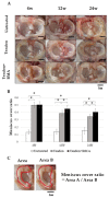
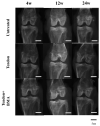
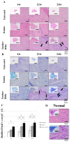
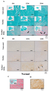
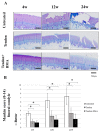
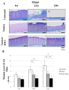
Bone marrow aspirate (BMA) is an excellent source of cells and growth factors and has been used successfully for bone, cartilage, and soft-tissue healing. This study aimed to investigate the histological and biomechanical properties of autogenous tendon graft by injecting BMA and its protective effect against degenerative changes in a rabbit model of meniscal defects. Adult white rabbits were divided into untreated, tendon, and tendon + BMA groups, and meniscal defects were created in the knees. The tendon graft and articular cartilage status were evaluated by macroscopic and histological analysis at 4, 12, and 24 weeks postoperatively among the three groups. The tendon graft in the tendon and tendon + BMA groups were used for biomechanical evaluation at 4, 12, and 24 weeks postoperatively. The meniscal covering ratio in the tendon + BMA group was better than that in the tendon and untreated groups at 12 and 24 weeks postoperatively. The matrix around the central portion of cells in the tendon + BMA group was positively stained by safranin O and toluidine blue staining with metachromasia at 24 weeks. The histological score of the tendon graft in the tendon + BMA group was significantly higher than that in the untreated and tendon groups at 12 and 24 weeks postoperatively. In the tendon + BMA group, cartilage erosion was not shown at 4 weeks, developed slowly, and was better preserved at 12 and 24 weeks compared to the untreated and tendon groups. Histological scores for the articular cartilage were significantly better in the tendon + BMA group at 24 weeks. The compressive stress on the tendon graft in the tendon + BMA group was significantly higher than that in the tendon group at 12 weeks postoperatively. Transplantation of autogenous tendon grafts by injecting BMA improved the histologic score of the regenerated meniscal tissue and was more effective than the tendon and untreated group for preventing cartilage degeneration in a rabbit model of massive meniscal defects.
Keywords: autogenous tendon graft; bone marrow aspirate; meniscus.
Conflict of interest statement
The authors declare no conflict of interest. The funders had no role in the design of the study; in the collection, analysis; or interpretation of data; in the writing of the manuscript; or in the decision to publish the results.
Figures


Similar articles
-
Transplantation of Parathyroid Hormone-Treated Achilles Tendon Promotes Meniscal Regeneration in a Rat Meniscal Defect Model.Am J Sports Med. 2022 Sep;50(11):3102-3111. doi: 10.1177/03635465221112954. Epub 2022 Aug 1. Am J Sports Med. 2022. PMID: 35914290
-
The effects of bone marrow or periosteum on tendon-to-bone tunnel healing in a rabbit model.Knee Surg Sports Traumatol Arthrosc. 2009 Feb;17(2):170-8. doi: 10.1007/s00167-008-0646-3. Epub 2008 Oct 22. Knee Surg Sports Traumatol Arthrosc. 2009. PMID: 18941736
-
Bone Marrow-Derived Fibrin Clots Stimulate Healing of a Knee Meniscal Defect in a Rabbit Model.Arthroscopy. 2023 Jul;39(7):1662-1670. doi: 10.1016/j.arthro.2022.12.013. Epub 2022 Dec 24. Arthroscopy. 2023. PMID: 36574822
-
The protective effects of meniscal transplantation on cartilage. An experimental study in sheep.J Bone Joint Surg Am. 2000 Jan;82(1):80-8. doi: 10.2106/00004623-200001000-00010. J Bone Joint Surg Am. 2000. PMID: 10653087
-
Meniscal substitutes--human experience.Scand J Med Sci Sports. 1999 Jun;9(3):146-57. doi: 10.1111/j.1600-0838.1999.tb00445.x. Scand J Med Sci Sports. 1999. PMID: 10380271 Review.
Cited by
-
Using Single Peroneal Longus Tendon Graft for Segmental Meniscus Transplantation and Revision Anterior Cruciate Ligament Combined Anterolateral Reconstruction.Medicina (Kaunas). 2023 Aug 21;59(8):1497. doi: 10.3390/medicina59081497. Medicina (Kaunas). 2023. PMID: 37629787 Free PMC article.
-
The Use of Free Fibula Flap in Different Extremities and Our Clinical Results.Cureus. 2023 Oct 22;15(10):e47450. doi: 10.7759/cureus.47450. eCollection 2023 Oct. Cureus. 2023. PMID: 37877106 Free PMC article.
References
MeSH terms
Substances
LinkOut - more resources
Full Text Sources

