Review
. 2022 Oct 19;23(20):12508.
doi: 10.3390/ijms232012508.

Molecular Dissection of TDP-43 as a Leading Cause of ALS/FTLD

Affiliations
Review

Molecular Dissection of TDP-43 as a Leading Cause of ALS/FTLD

Yoshitaka Tamaki et al. Int J Mol Sci. .

Abstract

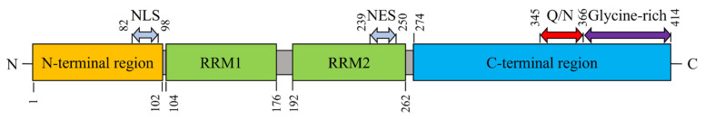
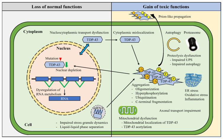
TAR DNA binding protein 43 (TDP-43) is a DNA/RNA binding protein involved in pivotal cellular functions, especially in RNA metabolism. Hyperphosphorylated and ubiquitinated TDP-43-positive neuronal cytoplasmic inclusions are identified in the brain and spinal cord in most cases of amyotrophic lateral sclerosis (ALS) and a substantial proportion of frontotemporal lobar degeneration (FTLD) cases. TDP-43 dysfunctions and cytoplasmic aggregation seem to be the central pathogenicity in ALS and FTLD. Therefore, unraveling both the physiological and pathological mechanisms of TDP-43 may enable the exploration of novel therapeutic strategies. This review highlights the current understanding of TDP-43 biology and pathology, describing the cellular processes involved in the pathogeneses of ALS and FTLD, such as post-translational modifications, RNA metabolism, liquid-liquid phase separation, proteolysis, and the potential prion-like propagation propensity of the TDP-43 inclusions.

Keywords: ALS; FTLD; RNA metabolism; TDP-43; aggregation; neurodegenerative diseases; phase separation.


Conflict of interest statement

The authors declare no conflict of interest.

Figures

Figure 1
Figure 1
The structure of TAR DNA-binding protein 43 (TDP-43) protein. TDP-43 is composed of an N-terminal region, two RNA recognition motifs (RRM1 and RRM2), a nuclear localization signal (NLS), a nuclear export signal (NES), and a C-terminal region (with a prion-like glutamine-/asparagine-rich [Q/N] region and a glycine-rich region). The numbers represent amino acid positions.
Figure 2
Figure 2
Molecular mechanisms underlying TDP-43 pathogenesis. Various mechanisms have been identified in the TDP-43 pathogenesis. The impairments of TDP-43 autoregulation and nucleocytoplasmic shuttling result in the dysregulation of RNA metabolism and increased cytoplasmic TDP-43 mislocalization. TDP-43 mislocalization triggers various post-translational modifications, including hyperphosphorylation, ubiquitination, acetylation, and C-terminal fragmentation, which facilitate TDP-43 oligomerization and aggregation. The FALS-linked mutations enhance intrinsic aggregation. The nuclear depletion and cytoplasmic aggregation of TDP-43 induce multiple cytotoxic effects, such as aberrant stress granule dynamics, liquid–liquid phase separation, mitochondrial dysfunction, endoplasmic reticulum (ER) stress, impaired axonal transport, and proteolysis dysfunction. TDP-43 aggregates show prion-like cell-to-cell propagation, which may confer the disease progression.
Figure 3
Figure 3
The distributions of TDP-43 pathology in the extra motor regions. The distribution of TDP-43 pathology in ALS-FTLD differs depending on the clinical types of cognitive impairments, such as: (A) executive dysfunction; (B) language dysfunction; (C) verbal fluency dysfunction; (D) behavioral dysfunction; and (E) semantic aphasia. Furthermore, (F) Limbic-predominant age-related TDP-43 encephalopathy (LATE) exhibits TDP-43 pathology in the limbic system, the frontal lobe, and the temporal lobe. The red areas show the distributions of TDP-43 pathology in the cerebrum.

References

    1. Mejzini R., Flynn L.L., Pitout I.L., Fletcher S., Wilton S.D., Akkari P.A. ALS genetics, mechanisms, and therapeutics: Where are we now? Front. Neurosci. 2019;13:1310. doi: 10.3389/fnins.2019.01310. - DOI - PMC - PubMed
    1. Bang J., Spina S., Miller B.L. Frontotemporal dementia. Lancet. 2015;386:1672–1682. doi: 10.1016/S0140-6736(15)00461-4. - DOI - PMC - PubMed
    1. Burrell J.R., Halliday G.M., Kril J.J., Ittner L.M., Götz J., Kiernan M.C., Hodges J.R. The frontotemporal dementia-motor neuron disease continuum. Lancet. 2016;388:919–931. doi: 10.1016/S0140-6736(16)00737-6. - DOI - PubMed
    1. Ringholz G.M., Appel S.H., Bradshaw M., Cooke N.A., Mosnik D.M., Schulz P.E. Prevalence and patterns of cognitive impairment in sporadic ALS. Neurology. 2005;65:586–590. doi: 10.1212/01.wnl.0000172911.39167.b6. - DOI - PubMed
    1. Arai T., Hasegawa M., Akiyama H., Ikeda K., Nonaka T., Mori H., Mann D., Tsuchiya K., Yoshida M., Hashizume Y., et al. TDP-43 is a component of ubiquitin-positive tau-negative inclusions in frontotemporal lobar degeneration and amyotrophic lateral sclerosis. Biochem. Biophys. Res. Commun. 2006;351:602–611. doi: 10.1016/j.bbrc.2006.10.093. - DOI - PubMed