

The Structural Interactions of Molecular and Fibrillar Collagen Type I with Fibronectin and Its Role in the Regulation of Mesenchymal Stem Cell Morphology and Functional Activity
- PMID: 36293432
- PMCID: PMC9604100
- DOI: 10.3390/ijms232012577
The Structural Interactions of Molecular and Fibrillar Collagen Type I with Fibronectin and Its Role in the Regulation of Mesenchymal Stem Cell Morphology and Functional Activity
Abstract
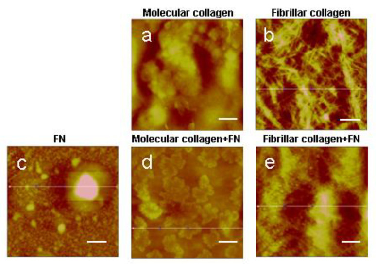
The observed differences in the structure of native tissue and tissue formed in vitro cause the loss of functional activity of cells cultured in vitro. The lack of fundamental knowledge about the protein mechanism interactions limits the ability to effectively create in vitro native tissue. Collagen is able to spontaneously assemble into fibrils in vitro, but in vivo, other proteins, for example fibronectin, have a noticeable effect on this process. The molecular or fibrillar structure of collagen plays an equally important role. Therefore, we studied the interaction of the molecular and fibrillar structure of collagen with fibronectin. Atomic force and transmission electron microscopy showed that the presence of fibronectin does not affect the native structure and diameter of collagen fibrils. Confocal microscopy demonstrated that the collagen structure affects the cell morphology. Cells are better spread on molecular collagen compared with cells cultured on fibrillar collagen. Fibronectin promotes the formation of a large number of focal contacts, while in combination with collagen of both forms, its effect is leveled. Thus, understanding the mechanisms of the relationship between the protein structure and composition will effectively manage the creation in vitro of a new tissue with native properties.
Keywords: actin cytoskeleton; cell focal contacts; fibronectin; molecular and fibrillar collagens.
Conflict of interest statement
The authors declare no competing financial interests.
Figures


Similar articles
-
Intracellular calreticulin regulates multiple steps in fibrillar collagen expression, trafficking, and processing into the extracellular matrix.J Biol Chem. 2010 Mar 5;285(10):7067-78. doi: 10.1074/jbc.M109.006841. Epub 2009 Dec 31. J Biol Chem. 2010. PMID: 20044481 Free PMC article.
-
Schwann cells use a novel collagen-dependent mechanism for fibronectin fibril assembly.J Cell Sci. 1998 Sep;111 ( Pt 18):2763-77. doi: 10.1242/jcs.111.18.2763. J Cell Sci. 1998. PMID: 9718369
-
Vascular smooth muscle cells orchestrate the assembly of type I collagen via alpha2beta1 integrin, RhoA, and fibronectin polymerization.Am J Pathol. 2003 Sep;163(3):1045-56. doi: 10.1016/s0002-9440(10)63464-5. Am J Pathol. 2003. PMID: 12937145 Free PMC article.
-
Fibrillar Collagens.Subcell Biochem. 2017;82:457-490. doi: 10.1007/978-3-319-49674-0_14. Subcell Biochem. 2017. PMID: 28101870 Review.
-
Collagen fibrillogenesis: fibronectin, integrins, and minor collagens as organizers and nucleators.Curr Opin Cell Biol. 2008 Oct;20(5):495-501. doi: 10.1016/j.ceb.2008.06.008. Epub 2008 Jul 30. Curr Opin Cell Biol. 2008. PMID: 18640274 Free PMC article. Review.
Cited by
-
Plasma Fibronectin as a Novel Predictor of Coronary Heart Disease: A Retrospective Study.J Cardiovasc Dev Dis. 2023 Oct 2;10(10):415. doi: 10.3390/jcdd10100415. J Cardiovasc Dev Dis. 2023. PMID: 37887862 Free PMC article.
-
Advanced In Vitro Three-Dimensional Skin Models of Atopic Dermatitis.Tissue Eng Regen Med. 2023 Jul;20(4):539-552. doi: 10.1007/s13770-023-00532-1. Epub 2023 Mar 30. Tissue Eng Regen Med. 2023. PMID: 36995643 Free PMC article. Review.
-
Epigallocatechin-3-gallate inhibits the collagen accumulation of oral submucous fibrosis induced by arecoline.Front Pharmacol. 2025 Jan 31;16:1540559. doi: 10.3389/fphar.2025.1540559. eCollection 2025. Front Pharmacol. 2025. PMID: 39959427 Free PMC article.
-
Focusing on the Native Matrix Proteins in Calcific Aortic Valve Stenosis.JACC Basic Transl Sci. 2023 Mar 29;8(8):1028-1039. doi: 10.1016/j.jacbts.2023.01.009. eCollection 2023 Aug. JACC Basic Transl Sci. 2023. PMID: 37719438 Free PMC article. Review.
-
BMP-Binding Polysulfonate Brushes to Control Growth Factor Presentation and Regulate Matrix Remodelling.ACS Appl Mater Interfaces. 2024 Aug 7;16(31):40455-40468. doi: 10.1021/acsami.4c05139. Epub 2024 Jul 29. ACS Appl Mater Interfaces. 2024. PMID: 39072446 Free PMC article.
References
MeSH terms
Substances
Grants and funding
LinkOut - more resources
Full Text Sources
