

Treadmill Exercise Reduces Neuroinflammation, Glial Cell Activation and Improves Synaptic Transmission in the Prefrontal Cortex in 3 × Tg-AD Mice
- PMID: 36293516
- PMCID: PMC9604030
- DOI: 10.3390/ijms232012655
Treadmill Exercise Reduces Neuroinflammation, Glial Cell Activation and Improves Synaptic Transmission in the Prefrontal Cortex in 3 × Tg-AD Mice
Abstract
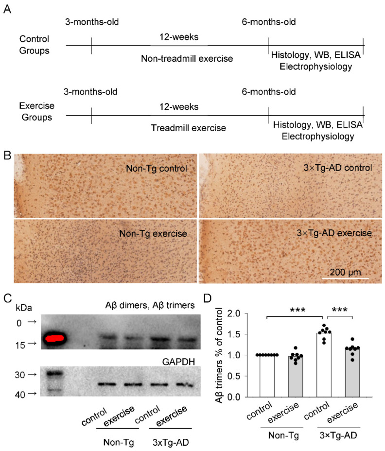
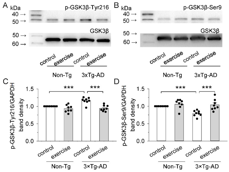
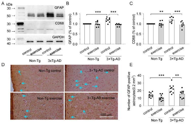
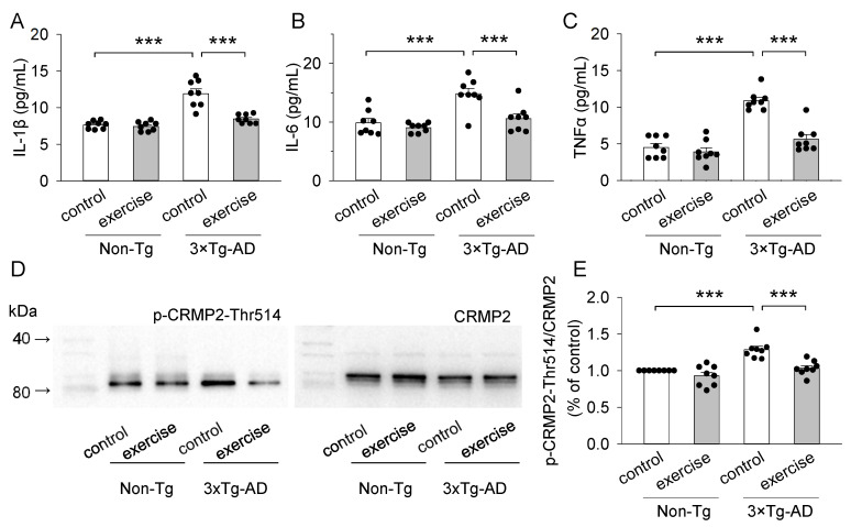
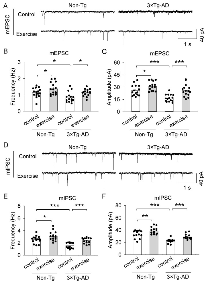
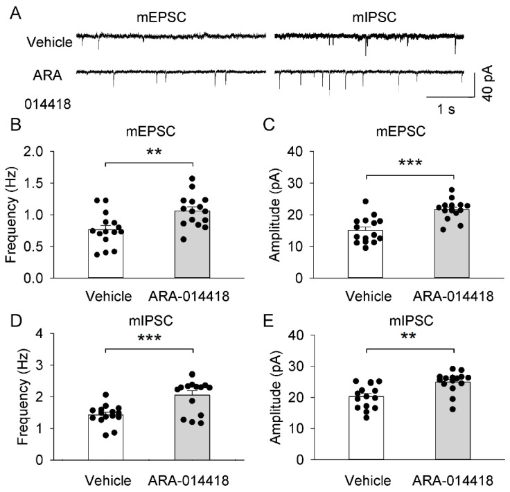
Physical exercise improves memory and cognition in physiological aging and Alzheimer's disease (AD), but the mechanisms remain poorly understood. Here, we test the hypothesis that Aβ oligomer accumulation, neuroinflammation, and glial cell activation may lead to disruption of synaptic transmission in the prefrontal cortex of 3 × Tg-AD Mice, resulting in impairment of learning and memory. On the other hand, treadmill exercise could prevent the pathogenesis and exert neuroprotective effects. Here, we used immunohistochemistry, western blotting, enzyme-linked immunosorbent assay, and slice electrophysiology to analyze the levels of GSK3β, Aβ oligomers (Aβ dimers and trimers), pro-inflammatory cytokines (IL-1β, IL-6, and TNFα), the phosphorylation of CRMP2 at Thr514, and synaptic currents in pyramidal neurons in the prefrontal cortex. We show that 12-week treadmill exercise beginning in three-month-old mice led to the inhibition of GSK3β kinase activity, decreases in the levels of Aβ oligomers, pro-inflammatory cytokines (IL-1β, IL-6, and TNFα), and the phosphorylation of CRMP2 at Thr514, reduction of microglial and astrocyte activation, and improvement of excitatory and inhibitory synaptic transmission of pyramidal neurons in the prefrontal cortex of 3 × Tg-AD Mice. Thus, treadmill exercise reduces neuroinflammation, glial cell activation and improves synaptic transmission in the prefrontal cortex in 3 × Tg-AD mice, possibly related to the inhibition of GSK3β kinase activity.
Keywords: 3 × Tg-AD mice; Aβ oligomer; GSK3β; exercise; glial cell; inflammatory factor; synaptic plasticity.
Conflict of interest statement
The authors declare no conflict of interest.
Figures
Similar articles
-
GSK3β is involved in promoting Alzheimer's disease pathologies following chronic systemic exposure to Porphyromonas gingivalis lipopolysaccharide in amyloid precursor proteinNL-F/NL-F knock-in mice.Brain Behav Immun. 2021 Nov;98:1-12. doi: 10.1016/j.bbi.2021.08.213. Epub 2021 Aug 13. Brain Behav Immun. 2021. PMID: 34391814 Free PMC article.
-
Chlorzoxazone exhibits neuroprotection against Alzheimer's disease by attenuating neuroinflammation and neurodegeneration in vitro and in vivo.Int Immunopharmacol. 2020 Nov;88:106790. doi: 10.1016/j.intimp.2020.106790. Epub 2020 Aug 11. Int Immunopharmacol. 2020. PMID: 32795892
-
The neuroprotective N-terminal amyloid-β core hexapeptide reverses reactive gliosis and gliotoxicity in Alzheimer's disease pathology models.J Neuroinflammation. 2023 May 27;20(1):129. doi: 10.1186/s12974-023-02807-9. J Neuroinflammation. 2023. PMID: 37245024 Free PMC article.
-
Treadmill Exercise Prevents Decline in Spatial Learning and Memory in 3×Tg-AD Mice through Enhancement of Structural Synaptic Plasticity of the Hippocampus and Prefrontal Cortex.Cells. 2022 Jan 12;11(2):244. doi: 10.3390/cells11020244. Cells. 2022. PMID: 35053360 Free PMC article.
-
Preventing synaptic deficits in Alzheimer's disease by inhibiting tumor necrosis factor alpha signaling.IBRO Rep. 2018 Feb 2;4:18-21. doi: 10.1016/j.ibror.2018.01.003. eCollection 2018 Jun. IBRO Rep. 2018. PMID: 30135948 Free PMC article. Review.
Cited by
-
GSK3-Driven Modulation of Inflammation and Tissue Integrity in the Animal Model.Int J Mol Sci. 2024 Jul 29;25(15):8263. doi: 10.3390/ijms25158263. Int J Mol Sci. 2024. PMID: 39125833 Free PMC article. Review.
-
Neuroinflammation-A Crucial Factor in the Pathophysiology of Depression-A Comprehensive Review.Biomolecules. 2025 Mar 30;15(4):502. doi: 10.3390/biom15040502. Biomolecules. 2025. PMID: 40305200 Free PMC article. Review.
-
Editorial: The impact of physical activity on white matter during healthy aging.Front Aging Neurosci. 2023 Feb 20;15:1140767. doi: 10.3389/fnagi.2023.1140767. eCollection 2023. Front Aging Neurosci. 2023. PMID: 36891554 Free PMC article. No abstract available.
-
Treadmill running on neuropathic pain: via modulation of neuroinflammation.Front Mol Neurosci. 2024 Jun 26;17:1345864. doi: 10.3389/fnmol.2024.1345864. eCollection 2024. Front Mol Neurosci. 2024. PMID: 38989156 Free PMC article. Review.
-
Exercise mimetics: a novel strategy to combat neuroinflammation and Alzheimer's disease.J Neuroinflammation. 2024 Feb 2;21(1):40. doi: 10.1186/s12974-024-03031-9. J Neuroinflammation. 2024. PMID: 38308368 Free PMC article. Review.
References
MeSH terms
Substances
Grants and funding
LinkOut - more resources
Full Text Sources
Medical
Molecular Biology Databases
Miscellaneous
