. 2022 Oct 21;23(20):12696.
doi: 10.3390/ijms232012696.

The Transcriptomic Landscape of Pediatric Astrocytoma

Affiliations

The Transcriptomic Landscape of Pediatric Astrocytoma

Abrahan Hernández-Hernández et al. Int J Mol Sci. .

Abstract

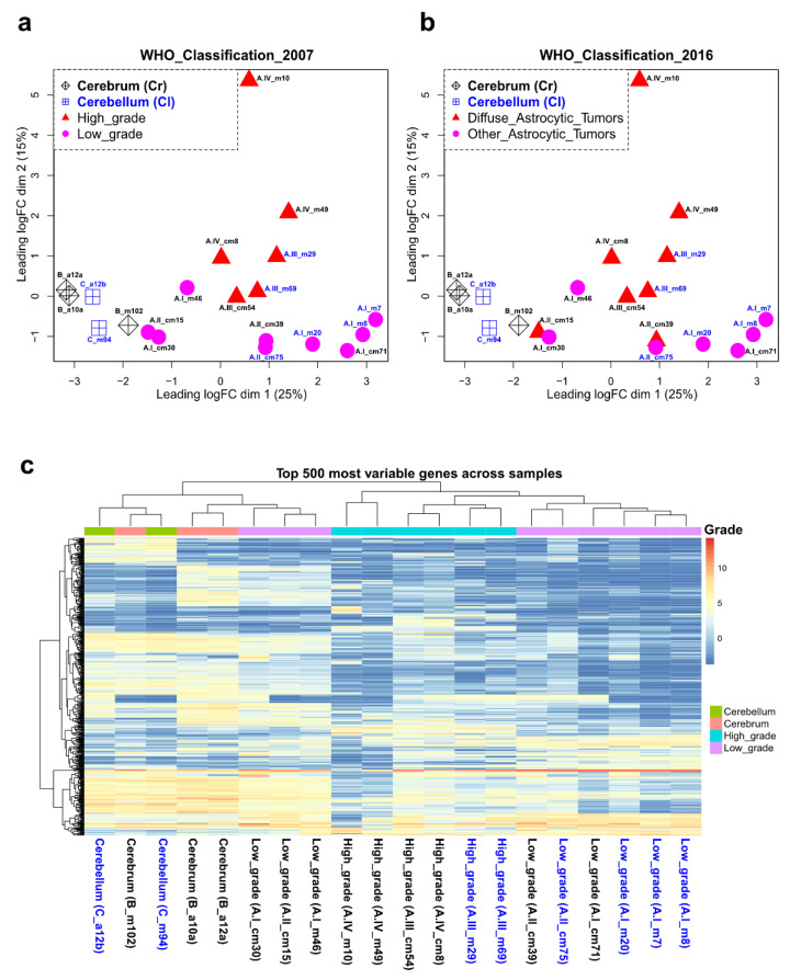
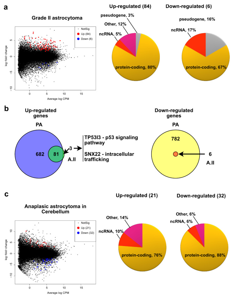
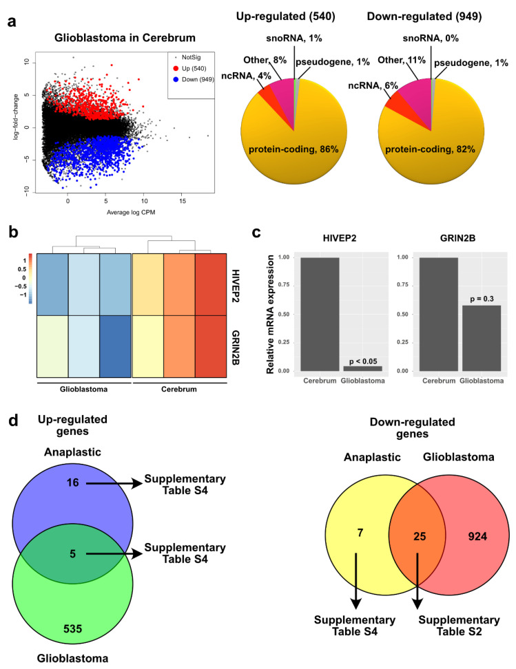
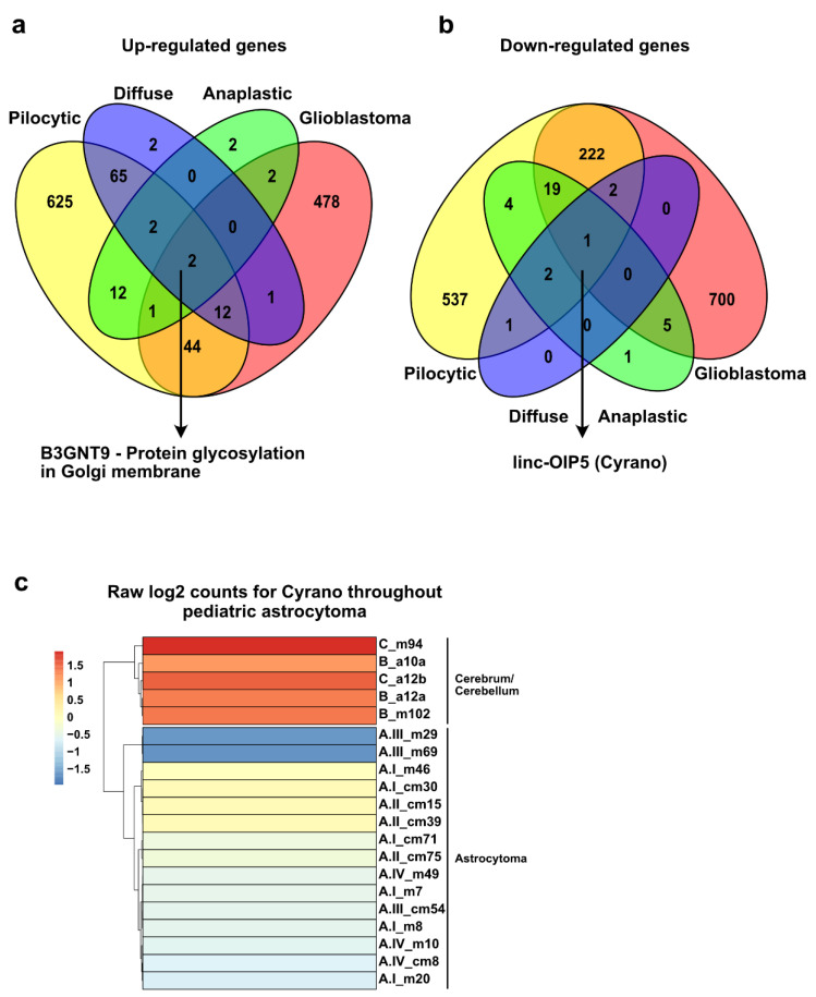
Central nervous system tumors are the most common solid neoplasia during childhood and represent one of the leading causes of cancer-related mortality. Tumors arising from astrocytic cells (astrocytomas) are the most frequently diagnosed, and according to their histological and pathological characteristics, they are classified into four categories. However, an additional layer of molecular classification considering the DNA sequence of the tumorigenesis-associated genes IDH1/2 and H3F3A has recently been incorporated into the classification guidelines. Although mutations in H3F3A are found exclusively in a subtype of grade IV pediatric astrocytoma, mutations in IDH1/2 genes are very rare in children under 14 years of age. The transcriptomic profiles of astrocytoma in adults and children have been extensively studied. However, there is scarce information on these profiles in pediatric populations considering the status of tumorigenesis-associated genes. Therefore, here we report the transcriptomic landscape of the four grades of pediatric astrocytoma by RNA sequencing. We found several well-documented biological functions associated with the misregulated genes in the four grades of astrocytoma, as well as additional biological pathways. Among the four grades of astrocytoma, we found shared misregulated genes that could have implications in tumorigenesis. Finally, we identified a transcriptional signature for almost all grades of astrocytoma that could be used as a transcription-based identification method.

Keywords: RNA-seq; pediatric astrocytoma; transcriptome.


Conflict of interest statement

The authors declare no conflicts of interest.

Figures

Figure 1
Figure 1
Experimental design of the study. Fifteen pediatric astrocytoma samples and five control tissues were processed for total RNA sequencing (RNA-seq). The bioinformatic pipeline for RNA-seq data analysis is shown.
Figure 2
Figure 2
Clustering of astrocytoma transcriptomes. Multidimensional scaling plots displaying the unsupervised principal component analysis of the pediatric astrocytoma RNA-seq data (transcriptomes) classified according to the 2007 WHO criteria (a) and 2016 WHO criteria (b). (c) Heat map displaying the unsupervised hierarchical clustering of the pediatric astrocytoma based on the 500 most variable genes across samples.
Figure 3
Figure 3
Differential expression of genes in pediatric low-grade astrocytoma. (a) Mean difference graphs highlighting the upregulated (red) and downregulated (blue) genes (FC > 1.5, FDR < 0.05) in pilocytic astrocytoma in the cerebellum compared to healthy matching tissue. In the right side of the panels, pie charts displaying the heterogeneity (gene type) of the up- and downregulated genes. (b) Structure of the fusion transcript BRAF-KIAA1549 detected in cerebellar PA, derived from the tandem duplication at location 7q34 (c). (d) Protein domains retained in the fusion protein BRAF-KIAA1549. (e) Prescence (Yes) or absence (No) of BRAF-KIAA1549 transcript in PA from different location in the brain.
Figure 4
Figure 4
Differential expression of genes in pediatric anaplastic astrocytoma. (a) Mean-difference plot highlighting the upregulated (red) and downregulated (blue) genes (FC > 1.5, FDR < 0.05) in grade II astrocytoma compared to healthy matching tissue. In the right side of the panel, pie charts displaying the heterogeneity (gene type) of the up- and downregulated genes. (b) Venn diagram showing overlap in the number of upregulated genes (left) and downregulated genes (right) for PA and grade II pediatric astrocytoma. (c) Mean-difference plot highlighting the up- and downregulated genes (FC > 1.5, FDR < 0.05) in anaplastic astrocytoma in the cerebellum compared to healthy matching tissue. In the right side of the panel, pie charts displaying the heterogeneity (gene type) of the up- and downregulated genes.
Figure 5
Figure 5
Differential expression of genes in pediatric glioblastoma. (a) Mean-difference plot highlighting the upregulated (red) and downregulated (blue) genes (FC > 1.5, FDR < 0.05) in glioblastoma in the cerebrum compared to healthy matching tissue. In the right side, pie charts displaying the heterogeneity (gene type) of the up- and downregulated genes. (b) Heat map showing the expression of two randomly picked genes in pediatric glioblastoma and healthy cerebrum from our RNA-seq data. Color bar displays the log count values. (c) Relative expression of the genes from panel b in five different samples of pediatric glioblastoma, evaluated by RT-qPCR. (d) Venn diagram showing overlap in the number of upregulated genes (left) and downregulated genes (right) for anaplastic astrocytoma and glioblastoma.
Figure 6
Figure 6
Shared misregulated genes among all the grades of pediatric astrocytoma. (a,b) Venn diagrams showing overlap in the number of up- (a) and downregulated genes (b) in the four grades of pediatric astrocytoma compared to healthy matching tissue. (c) Expression of the lncRNA Cyrano throughout pediatric astrocytoma and healthy cerebellum/cerebrum. Color bar displays the raw log2 counts for Cyrano in our RNA-seq data.
Figure 7
Figure 7
Uniquely expressed genes in pediatric astrocytoma. (ad) Mean-difference plots highlighting the upregulated (red) and downregulated (blue) genes in pilocytic astrocytoma in the cerebellum (a) (FC > 1.1, FDR < 0.05), anaplastic astrocytoma in the cerebellum (b) (FC > 3, FDR < 0.05), glioblastoma in the cerebrum (c) (FC > 3.5, FDR < 0.05), and in healthy cerebrum and cerebellum (d) (FC > 5, FDR < 0.05). Comparisons were made with all the remaining grades of astrocytoma and healthy control tissue.
Figure 8
Figure 8
Clustering of pediatric astrocytomas according to their uniquely expressed markers. (a) Heat map displaying the hierarchical clustering of low- and high-grade pediatric astrocytoma and healthy cerebrum/cerebellum based on a panel of uniquely expressed genes. (b) Heatmap with hierarchical clustering of the ssGSEA enrichment scores for the genes in the transcriptional signature projected in the publicly available GEO dataset GSE73066 with the transcriptional profile of 47 pediatric PAs in different locations of the brain.

References

    1. Chico-Ponce de Leon F., Castro-Sierra E., Perezpena-Diazconti M., Gordillo-Dominguez L.F., Santana-Montero B.L., Rocha-Rivero L.E., Vaca-Ruiz M.E., Ríos-Alanís M., Sánchez-Herrera F., Valdés-Ordoño R. Pediatric Intracranial Tumors. Bol. Med. Hosp. Infant. Mex. 2016;63:367–381.
    1. Bhatia A., Pruthi S. Pediatric Brain Tumors: A Different Ball Game. Semin. Roentgenol. 2018;53:77–100. doi: 10.1053/j.ro.2017.11.007. - DOI - PubMed
    1. Pollack I.F., Agnihotri S., Broniscer A. Childhood Brain Tumors: Current Management, Biological Insights, and Future Directions. J. Neurosurg. Pediatr. 2019;23:261–273. doi: 10.3171/2018.10.PEDS18377. - DOI - PMC - PubMed
    1. Louis D.N., Perry A., Reifenberger G., von Deimling A., Figarella-Branger D., Cavenee W.K., Ohgaki H., Wiestler O.D., Kleihues P., Ellison D.W. The 2016 World Health Organization Classification of Tumors of the Central Nervous System: A Summary. Acta Neuropathol. 2016;131:803–820. doi: 10.1007/s00401-016-1545-1. - DOI - PubMed
    1. PDQ Pediatric Treatment Editorial Board . PDQ Cancer Information Summaries. National Cancer Institute (US); Bethesda, MD, USA: 2002. Childhood Astrocytomas Treatment (PDQ(R)): Health Professional Version. - PubMed