. 2022 Sep 21;8(10):991.
doi: 10.3390/jof8100991.

Reactivation of Coccidioidomycosis in a Mouse Model of Asymptomatic Controlled Disease

Affiliations

Reactivation of Coccidioidomycosis in a Mouse Model of Asymptomatic Controlled Disease

Lisa F Shubitz et al. J Fungi (Basel). .

Abstract

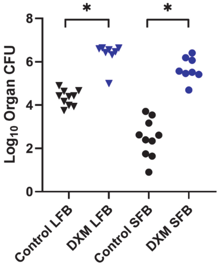
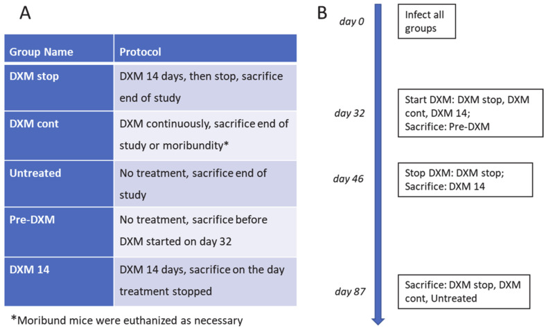
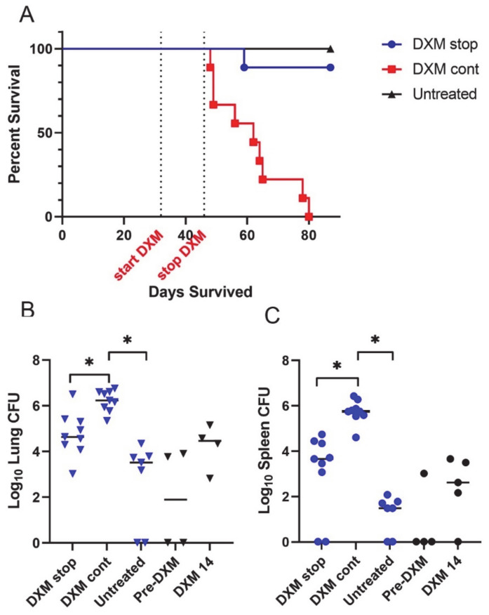
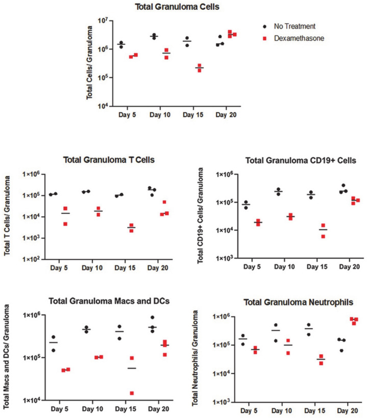
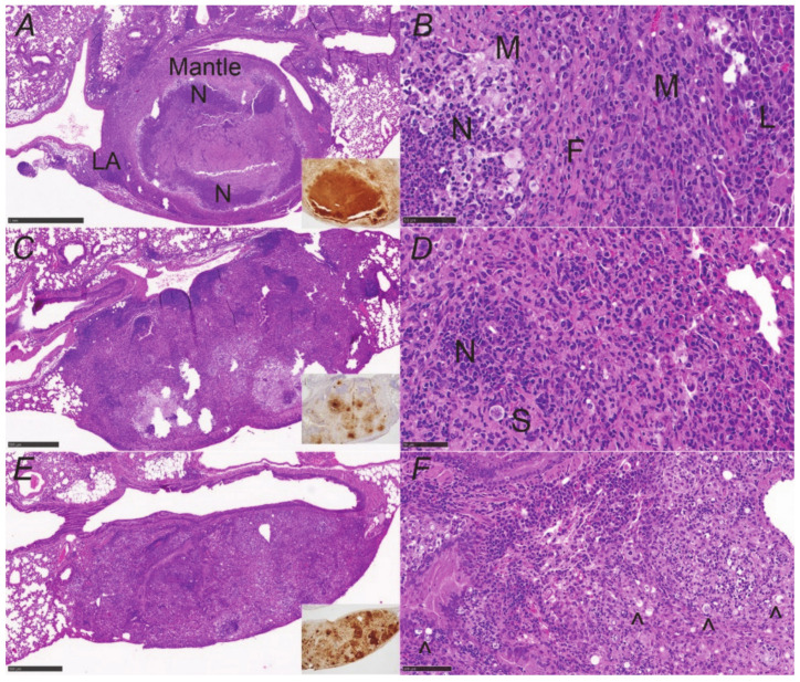
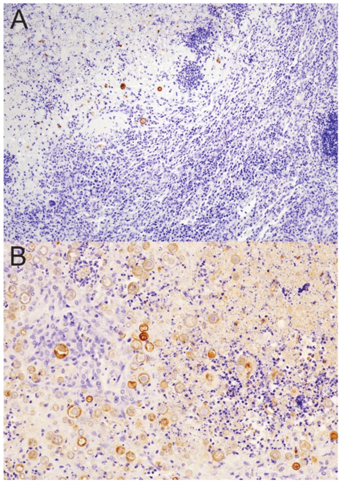
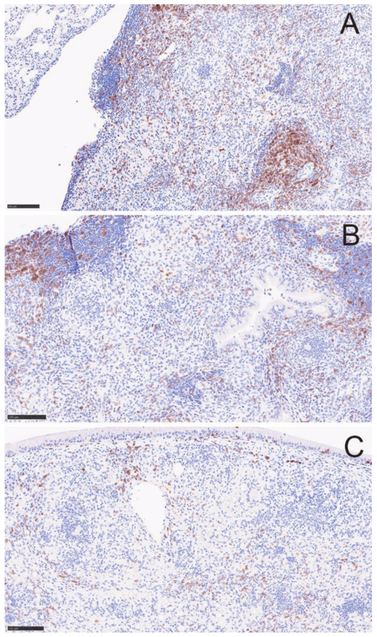
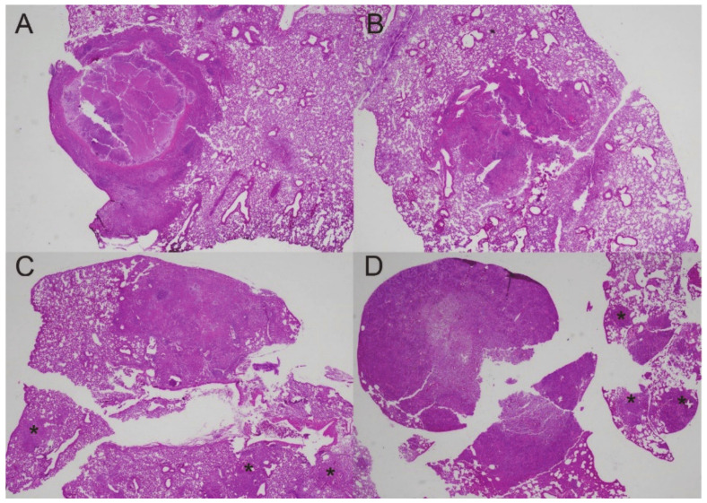
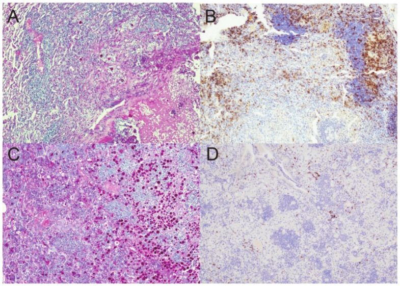
The majority of human coccidioidomycosis infections are asymptomatic or self-limited but may have sequestered spherules in highly structured granulomas. Under immunosuppression, reactivation of fungal growth can result in severe disease. B6D2F1 mice asymptomatically infected with C. posadasii strain 1038 were immunosuppressed with dexamethasone (DXM) in drinking water. Treated mice died 16−25 days later, while untreated mice survived (p < 0.001). Flow cytometry of lung granulomas on days 5, 10, 15, and 20 of DXM treatment showed immune cell populations decreased 0.5−1 log compared with untreated mice though neutrophils and CD19+IgD−IgM− cells rebounded by day 20. Histopathology demonstrated loss of granuloma structure by day 5 and increasing spherules through day 20. On day 20, T-cells were nearly absent and disorganized pyogranulomatous lesions included sheets of plasma cells and innumerable spherules. Mice given DXM for 14 days then stopped (DXM stop) survived 6 weeks (9/10). Lung fungal burdens were significantly lower (p = 0.0447) than mice that continued treatment (DXM cont) but higher than untreated mice. Histopathologically, DXM stop mice did not redevelop controlled granulomas by sacrifice, though T-cells were densely scattered throughout the lesions. This demonstrates a mouse model suitable for further study to understand the immunologic components responsible for maintenance control of coccidioidomycosis.

Keywords: Coccidioides; granuloma; immunosuppression; mice; reactivation.


Conflict of interest statement

None of the authors has a conflict of interest.

Figures

Figure 1
Figure 1
Organ fungal burdens of DXM treated and untreated mice. B6D2F1 mice (n = 8/group) were infected with 50 spores of Cp1038, and lung and spleen fungal burdens quantitated at termination after treatment with DXM in drinking water or no treatment. Fungal burdens were significantly higher in both lungs (LFB) and spleens (SFB) following continuous treatment with DXM compared with the untreated controls (p < 0.0001, both comparisons). (* Indicates statistical significance; statistical analysis—Kruskal–Wallis.).
Figure 2
Figure 2
Treatment and sacrifice protocol for comparison of mice treated for two weeks or continuously with DXM, 6 mg/L, in drinking water. Groups and treatment conditions (A) and timeline of study (B) for 2-week and continuous administration of DXM with right lung fungal burden and left lung histopathology.
Figure 3
Figure 3
Survival and fungal burdens of 2-week or continuous treatment with DXM. (A) Mice (n = 10/group) treated continuously with DXM (DXM cont) became moribund before scheduled sacrifice (day 87 post-infection). (B) Right lung fungal burdens of mice treated with DXM for 14 days and then discontinued (DXM stop) were intermediate compared with mice treated continuously (DXM cont) and untreated mice. DXM cont fungal burdens were significantly higher than the untreated (p < 0.0001) and DXM stop mice (p = 0.0447). The right lung fungal burdens of mice before the start of DXM (pre-DXM) and on the 14th day of treatment (DXM 14) show that the fungal burden increased rapidly with immunosuppression and remained approximately the same 6 weeks after discontinuation of DXM. (C) Spleen fungal burdens paralleled lung data (DXM stop vs. DXM cont, p = 0.0105; untreated vs. DXM cont, p = 0.0001). (* Indicates statistical significance. Statistical analysis: survival—Mann–Whitney U; organ fungal burdens—Kruskal–Wallis).
Figure 4
Figure 4
Flow cytometry counts of immune cells in treated and untreated mice. Total immune cells in the granulomas reduce between day 5 and 15 after starting DXM. There is a rebound on day 20. T-cell populations remain depressed at all time points, along with the modest reduction in macrophage/DC lines, but CD19+ B-lineage cells and neutrophils show an increase on day 20. Population subsets are shown in Supplementary Figures S1 and S2.
Figure 5
Figure 5
Granuloma destruction over time with DXM suppression. (A) Structure of a controlled granuloma showing it is well-demarcated from the surrounding lung and consists of a necrotic center with many neutrophils (N) and a mantle region composed of macrophages, fibroblasts, scattered neutrophils, and scattered lymphocytes (inset, neutrophil stain). Lymphoid aggregates (LA) are common on the periphery of the margin. (B) Higher-power image of (A) highlighting layers; F = fibroblasts, M = macrophages, L = lymphocytes, and N = neutrophils. (C) Day 5 of DXM administration shows a huge reduction in neutrophils in the core of the lesion (inset, neutrophil stain) and (D) is a higher-power image from the right side of the periphery with a large spherule (S) and a cluster of neutrophils (N) near the margins of the lesion. (E) Day 20 lesion showing complete loss of granuloma structure characterized by mixed inflammation of macrophages and resurgent neutrophils (inset, neutrophil stain) (pyogranulomatous lesion) with innumerable spherules throughout. (F) High-power image of (E) with some of the abundant spherules marked (black carat). Sheets of plasma cells are the main lymphocytic cells present. (H&E stain; insets—NIMP-R14 neutrophil stain. Magnification: (A,C,E)—×40; (B,D,F)—×200).
Figure 6
Figure 6
Coccidioides-specific stain of spherules prior to and after DXM immunosuppression. (A) There are few spherules (brown stain) confined to the necrotic core of a controlled granuloma in an untreated mouse. (B) Sheets of spherules are found throughout the disorganized pyogranulomatous lesion (former granuloma) in a mouse treated for 20 days with DXM. Note that many of the spherules in this image are only minimally stained. (Stain: polyclonal goat anti-PRA Ab with a hematoxylin counterstain; magnification (A)—×100; (B)—×200).
Figure 7
Figure 7
T-cells diminish over 20 days in DXM treated mice. CD3+ T-cells diminish in the lesions over time. (A) before treatment, (B) DXM Day 5, and (C) DXM day 20. Images are from the same lesions as in Figure 3 and further demonstrate the loss of the lymphoid aggregates. (Stain—anti-CD3 (brown) with hematoxylin counterstain; magnification ×100).
Figure 8
Figure 8
Granulomas at different time points before and after DXM suppression. (A) Controlled granuloma in untreated mouse; (B) after 14 days of DXM, the structure of the lesion is lost; (C) 6 weeks after stopping DXM, the pyogranulomatous lesion has not recovered the structure seen in (A), and there are additional centers of pyogranulomatous inflammation in the lung lobe (asterisks); (D) in a mouse treated continuously with DXM, the primary lesion on the left side of the image is intensely neutrophilic with innumerable spherules, and there are multiple additional lesions with many spherules beyond the large initial one (asterisks). (Stain—HE; magnification ×20).
Figure 9
Figure 9
Spherules and T-cells in DXM stop and DXM cont mice. Spherules (A) were few and T-cells (B) abundant throughout the pyogranulomatous lesions of mice 6 weeks after discontinuing a 14-day course of DXM. By contrast, spherules were numerous (C) and T-cells nearly absent (D) in mice receiving DXM continuously. (Stain—(A,C)—periodic acid Schiff stains spherules dark pink; (B,D)—anti-CD3 IHC stains the T-cells brown; magnification ×100).

Similar articles

Cited by

References

    1. Donovan F.M., Shubitz L., Powell D., Orbach M., Frelinger J., Galgiani J.N. Early Events in Coccidioidomycosis. Clin. Microbiol. Rev. 2019;33:e112-19. doi: 10.1128/CMR.00112-19. - DOI - PMC - PubMed
    1. Straub M., Schwartz L.W. Primary pulmonary arrested lesions of coccidioidomycosis and histoplasmosis. Am. J. Clin. Pathol. 1956;26:998–1009. doi: 10.1093/ajcp/26.9.998. - DOI - PubMed
    1. Sachdeva M., Ronaghi R., Mills P.K., Peterson M.W. Complications and Yield of Computed Tomography-Guided Transthoracic Core Needle Biopsy of Lung Nodules at a High-Volume Academic Center in an Endemic Coccidioidomycosis Area. Lung. 2016;194:379–385. doi: 10.1007/s00408-016-9866-3. - DOI - PubMed
    1. Deresinski S.C., Stevens D.A. Coccidioidomycosis in compromised hosts. Experience at Stanford University Hospital. Medicine. 1975;54:377–395. doi: 10.1097/00005792-197509000-00002. - DOI - PubMed
    1. Logan J.L., Blair J.E., Galgiani J.N. Coccidioidomycosis complicating solid organ transplantation. Semin. Respir. Infect. 2001;16:251–256. doi: 10.1053/srin.2001.29318. - DOI - PubMed

LinkOut - more resources